anterior half broadly conical while posterior half narrower and obliquely truncate
peristome runs obliquely from anterior to posterior
cytostome is accompanied on the right by a perizonal stripe of kineties
oral aperture located posteriorly
macronucleus spherical with an adjacent micronucleus
aggregate of brownish or yellowish granules anteriorly
symbiotic bacteria are present
contractile vacuole terminally
tuft of longer caudal cilia
inconspicuous extrusomes
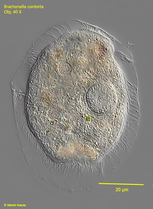
Brachonella contorta
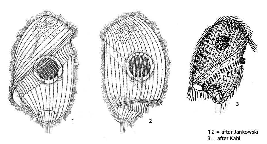
Brachonella contorta occurs in practically all my sampling locations and is very common. Originally this species was assigned by Kahl to the genus Metopus. Only in 1964 Jankowsi established the genus Brachonella. This genus differs from Metopus in having a posterior end broader than the anterior end and by a spiral peristome around the body axis. Brachonella contorta is described by Kahl (as Brachonella spiralis) as very coverslip sensitive. I can confirm this observation, but have found that some specimens can be fixed comparatively well. It was not obvious to me what distinguished these “stable” specimens from the others.
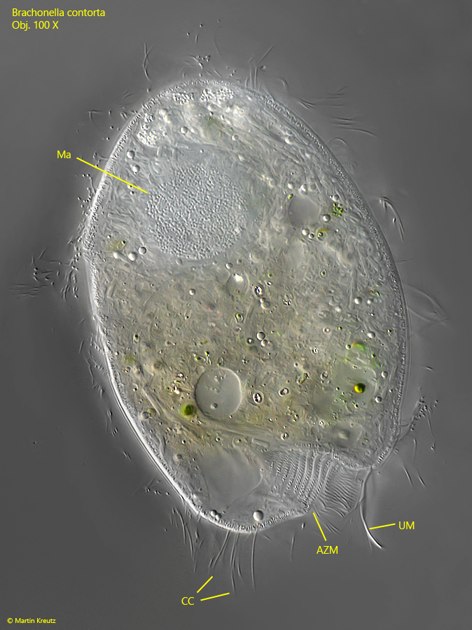
Fig. 1:Brachonella contorta. L = 105 µm. Ventral view of a freely swimming specimen. Obj. 100 X.
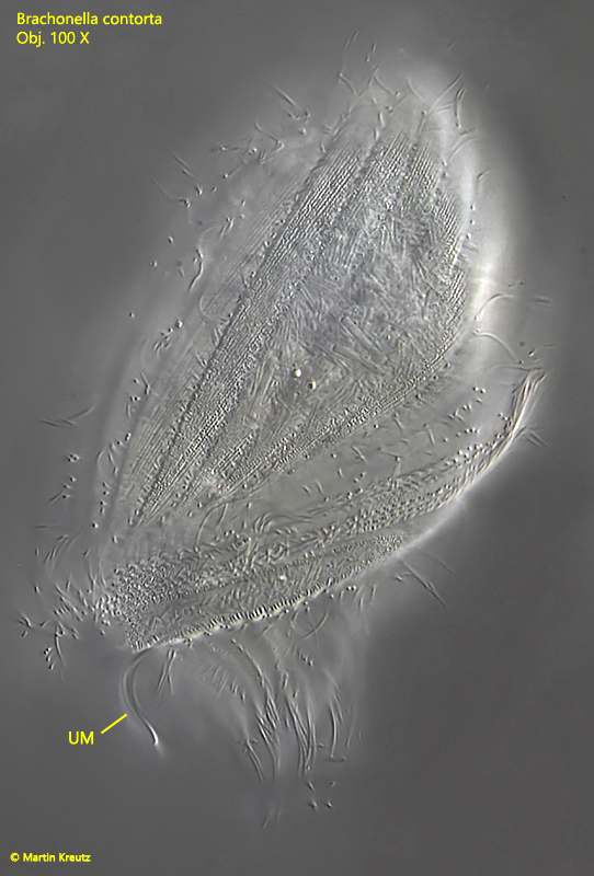
Fig. 2:Brachonella contorta. L = 130 µm. View from the right side. UM = undulating membranelle. Obj. 100 X.
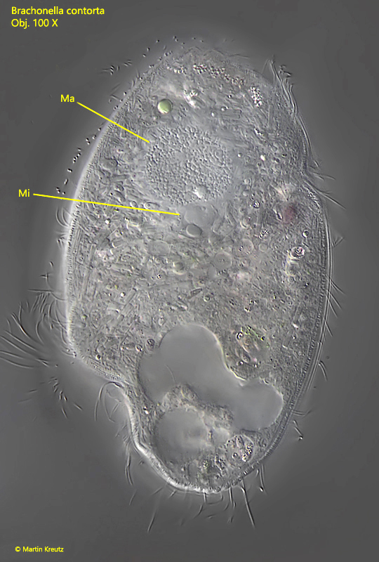
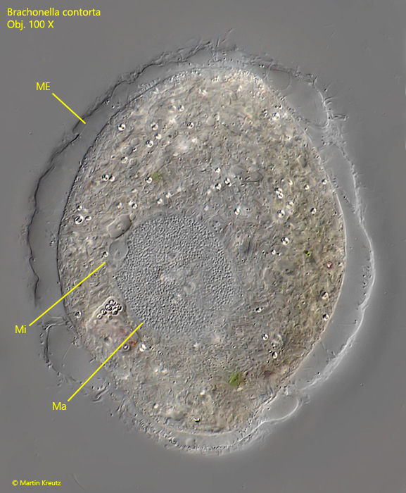
Fig. 3:Brachonella contorta. L = 105 µm. Ventral view with focal plane on the macronuclus (Ma) and the adjacent micronucleus (Mi) in the anterior half. Obj. 100 X.
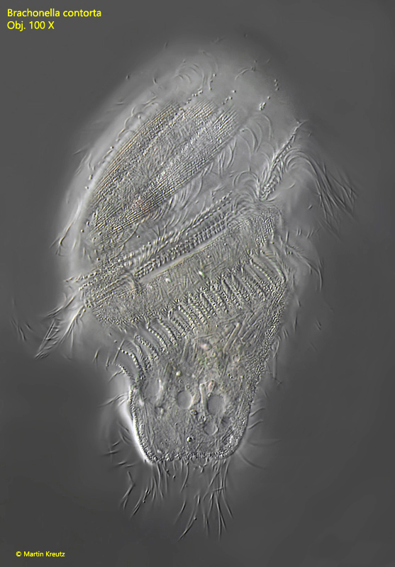
Fig. 4:Brachonella contorta. L = 110 µm. Dorsal view with focal plane on the adoral zone of membranelles (AZM) at the posterior end leading to the mouth opening and the undulatuing membrane (UM). CC = caudal cilia, Ma = macronucleus. Obj. 100 X.
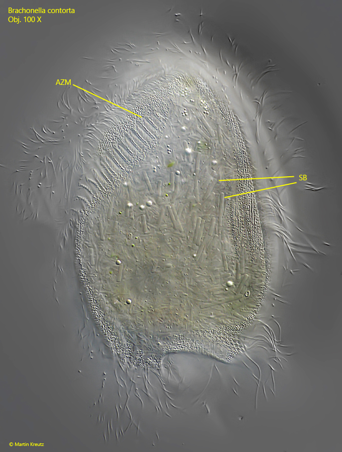
Fig. 5:Brachonella contorta. L = 115 µm. Dorsal view of the same specimen shown in fig. 4 with focal plane on the symbiotic bacteria (SB) in the cytoplasm and on the adoral zone of membranelles (AZM) beginning on the dorsal side. Obj. 100 X.
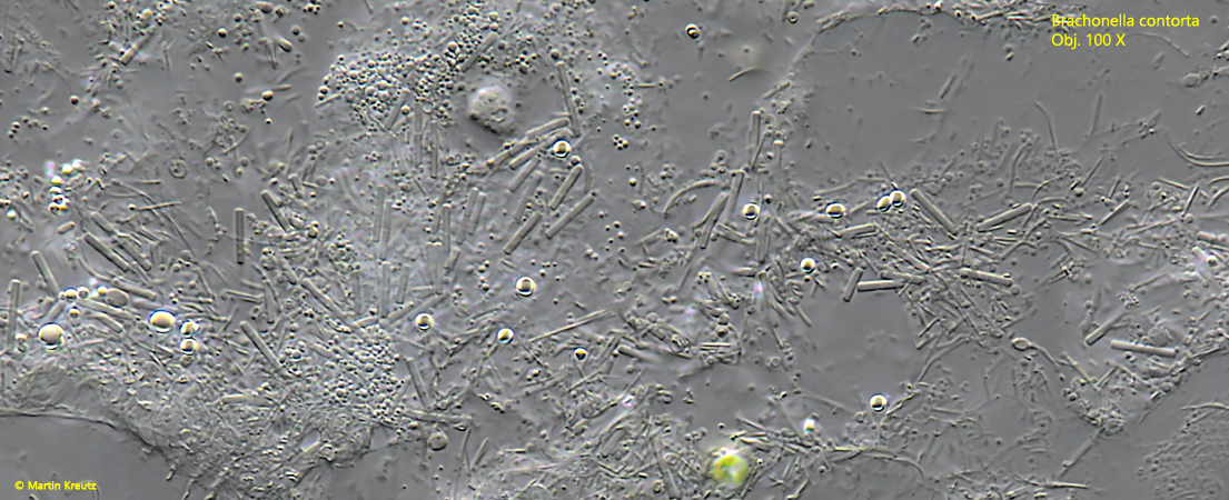
Fig. 6:Brachonella contorta. The scattered symbiotic bacteria in a squashed specimen. Obj. 100 X.
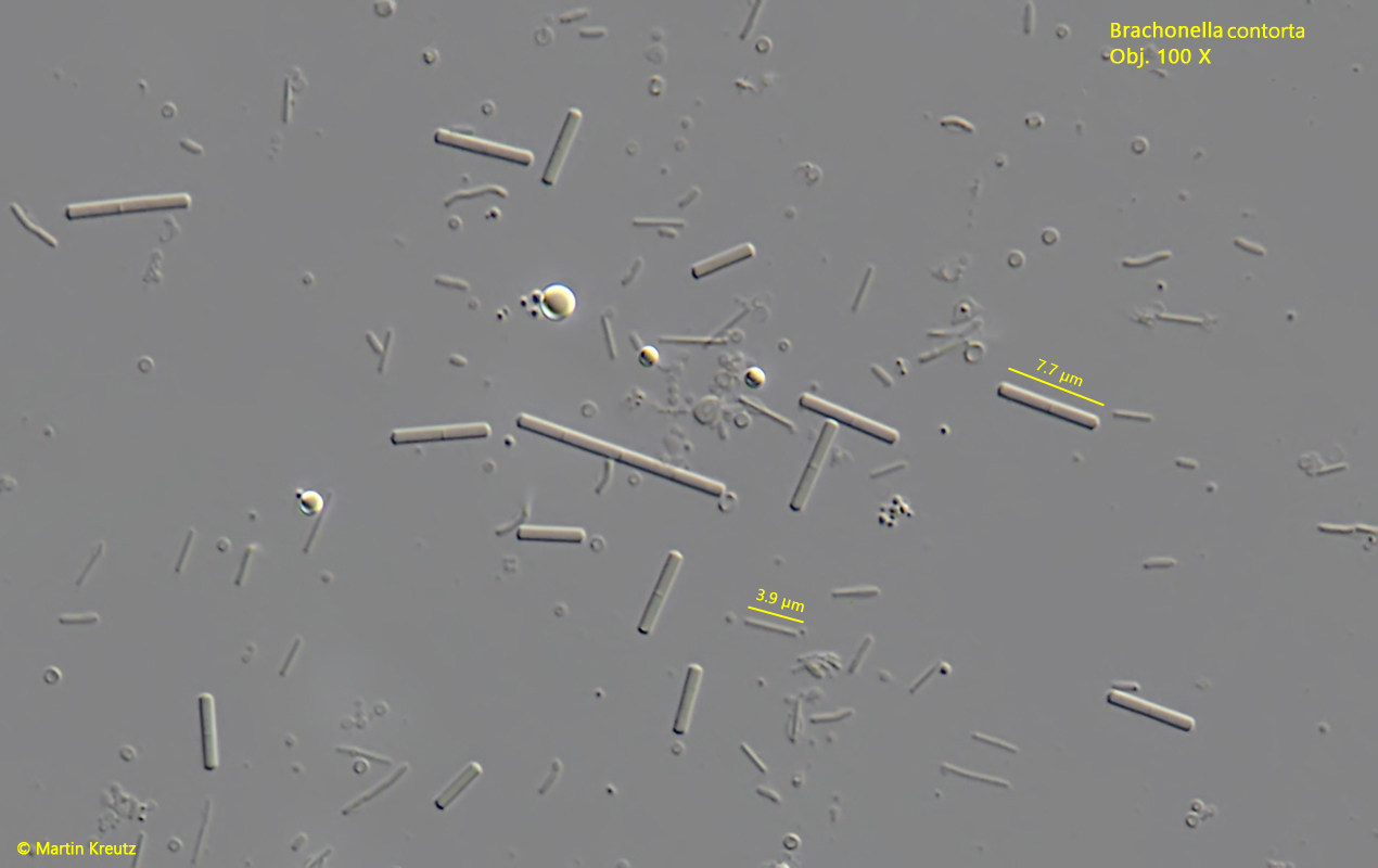
Fig. 7:Brachonella contorta. The symbiotic bacteria in a strongly squashed specimen. There are at least two different types of bacteria. The larger ones are about 5 µm long and oblong. The smaller ones are thin rods with a length of about 4 µm. Obj. 100 X.
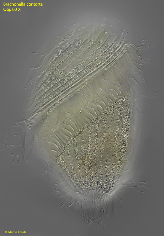
Fig. 8:Brachonella contorta. L = 130 µm. Ventral view of a second, larger specimen with a distinctly yellowish color. Obj. 60 X.
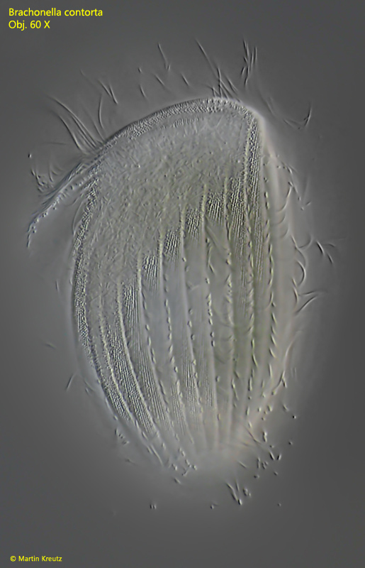
Fig. 9:Brachonella contorta. L = 130 µm. Dorsal view of a second specimen with a yellowish color. Obj. 60 X.
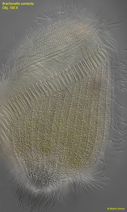
Fig. 10:Brachonella contorta. L = 130 µm. Detailed ventral view of a second specimen with a yellowish color. Obj. 100 X.
This yellowish specimen lacked the larger symbiotic bacteria (s. Fig. 10). This could be an indication of different compositions of symbiontic bacteria depending on the habitat.
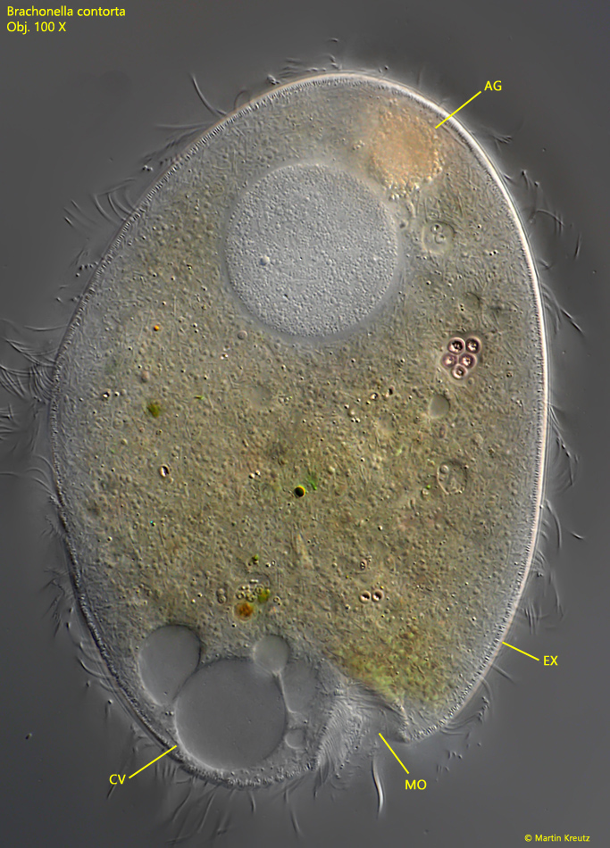
Fig. 11:Brachonella contorta. L = 130 µm. Focal plane on the macronucleus of a seond, yellowish colored specimen. In this specimen the larger symbiotic bacteria are not present. AG = anterior aggregation of granules, CV = contractile vacuole, EX = extrusomes, MO = mouth opening. Obj. 100 X.
Not all specimens of Brachonella contorta could be examined in as much detail as the specimens shown above. Most specimens burst already after careful placement of the coverslip. Whether this reaction is caused by the hydrostatic generated pressure of the coverslip or by the reduced “vertical freedom of movement” is hard to say exactly. At least in most cases it leads to the extrusomes (mucocysts) being abruptly ejected with simultaneous shrinkage and denaturation of the cell body and forming a kind of mucuous envelope around the ciliate (s. fig. 11). This self-destructive reaction is in my opinion in no case a beginning encystment. Other ciliates with extrusomes and/or mucocysts can expel them locally over a limited area and usually continue swimming afterwards (e.g. Paramecium caudatum or Frontonia leucas). Why such a self-destructive reaction starts in Brachonella (and especially in the genus Metopus) remains enigmatic for the time being.
Fig. 12:Brachonella contorta. A denatured specimen after the coverslip has been applied. Obj. 100 X.
Fig. 13: Brachonella spiralis. A squashed denatured specimen in detail. Ma = macronucleus, ME = mucuous envelope, Mi = micronucleus. Obj. 100 X.
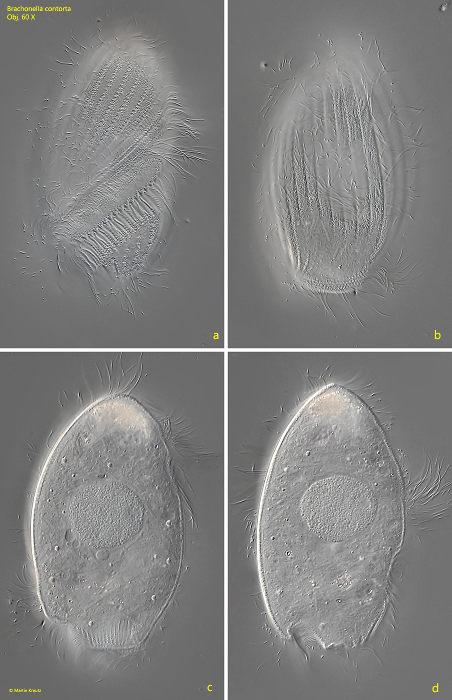
Fig. 14 a-d:Brachonella contorta. L = 110 µm. A freely swimming, almost colorless specimen from ventral (a), dorsal (b, c) and right (d). Obj. 60 X.