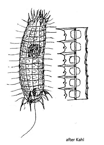
two D-shaped “windows” each mirror-symmetric to the main bar
one caudal cilium
macronucleus spherical in mid-body
apical mouth opening with basket of pharyngeal trichites
contractile vacuole subterminal
Pinacocoleps incurvus
Pinacocoleps incurvus was first described by Ehrenberg in 1833 as Coleps incurvus. Kahl later adopted this name. In 2008 Foissner et al. reexamined the species in the genus Coleps and transferred all species whose armour consists of 6 layers of plates per half cell (s. fig. 4 a) to the genus Pinacocoleps, which was created by Diesing in 1865.
Pinacocoleps incurvus is common in practically all my sites. The species can be recognized by the slender, almost curved body shape. This is due to the fact that one side of the body is flattened while the opposite side is convex. There is only one caudal cilium. However, the definite identification can only be made by examining the “windows” and their shape and arrangement in the armour. In Pinacocoleps incurvus they are D-shaped and are arranged in in mirror symmetric way opposite each other along the longitudinal main bars, as Kahl has drawn it (s. above).
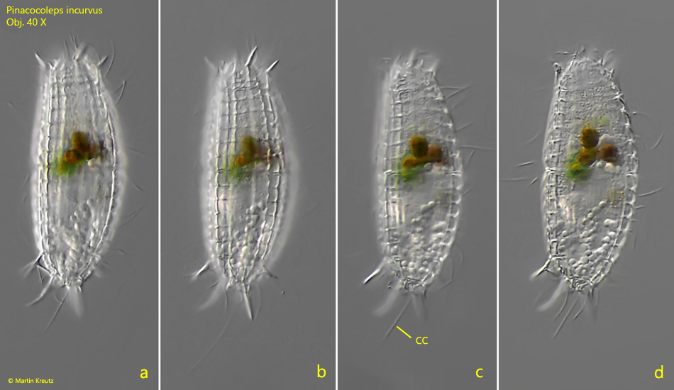
Fig. 1 a-d:Pinacocoleps incurvus. L = 76 µm. A freely swimming specimen. CC = caudal cilium. Obj. 40 X.
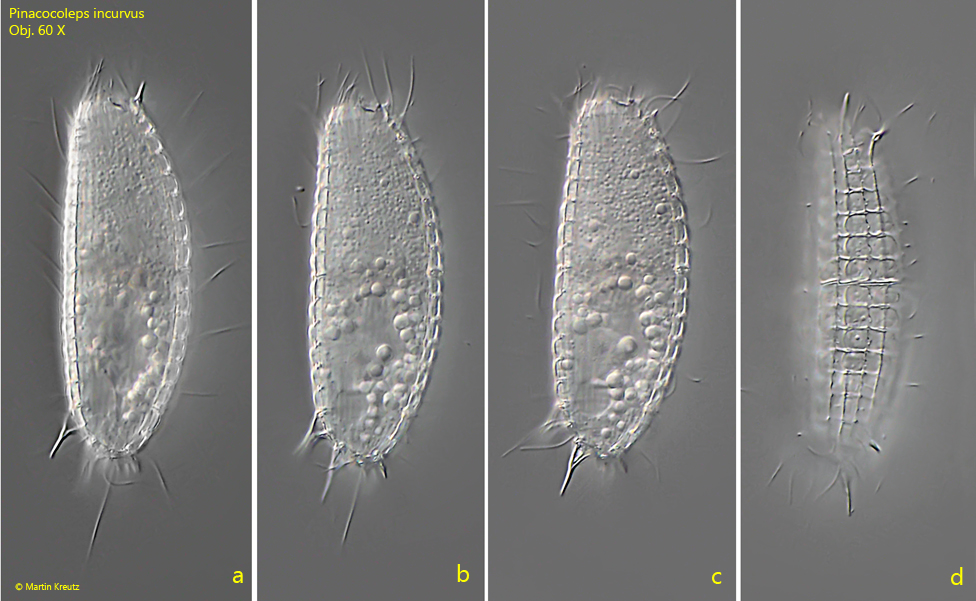
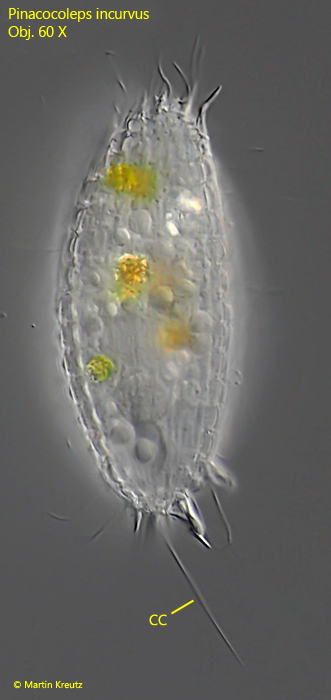
Fig. 2 a-d:Pinacocoleps incurvus. L = 60 µm. A second freely swimming specimen. Obj. 60 X.
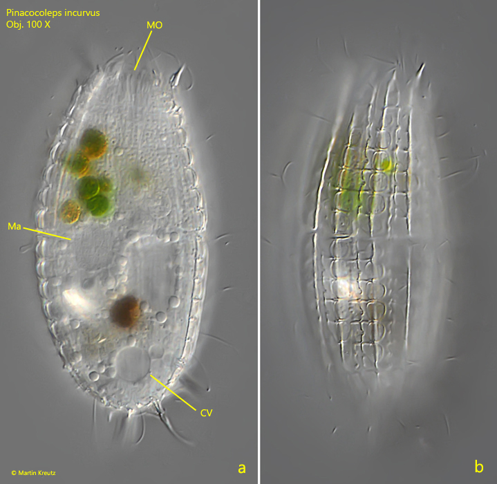
Fig. 3 a-b:Pinacocoleps incurvus. L = 76 µm. The slightly squashed specimen shown in fig. 1 a-e. CV = contractile vacuole, Ma = macronucleus, MO = mouth opening. Obj. 100 X.
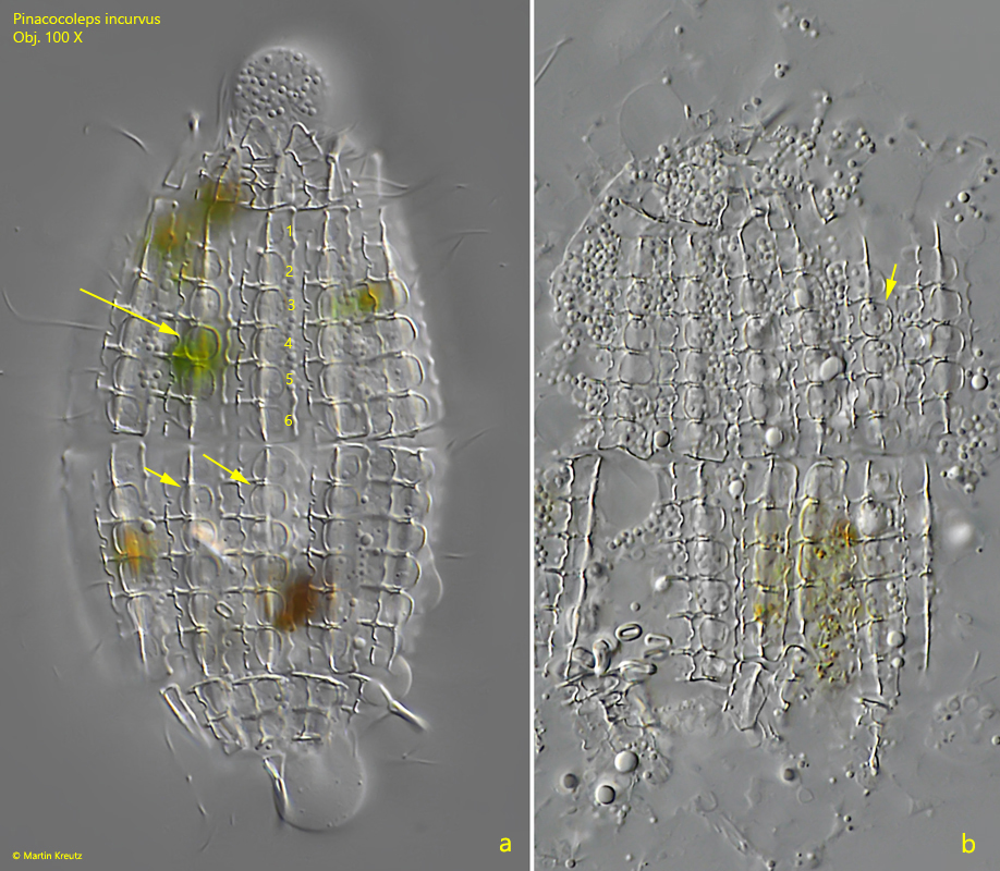
Fig. 4 a-b:Pinacocoleps incurvus. The squashed specimen shown in fig. 1 a-e. Note the D-shaped “windows” each mirror-symmetric to the main bar (arrows). Per half cell each 6 “windows” are arranged in a row (1–6). Obj. 100 X.
Fig. 5:Pinacocoleps incurvus. L = 73 µm. A second freely swimming specimen. CC = caudal cilium. Obj. 60 X.
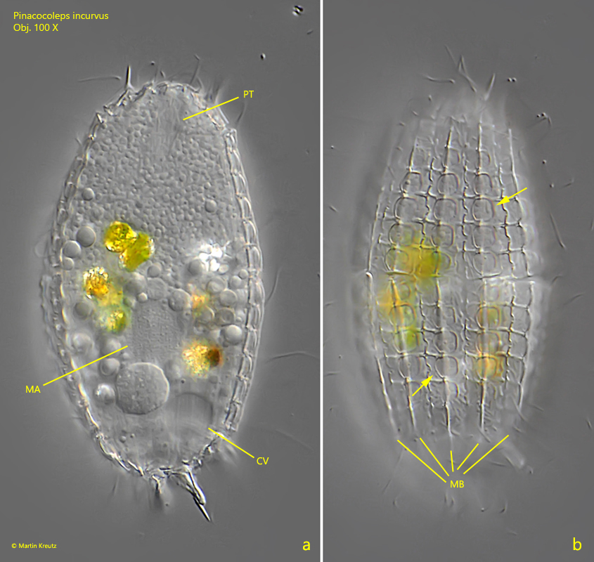
Fig. 6 a-b:Pinacocoleps incurvus. The squashed specimen shown in fig. 4. The D-shaped “windows” (arrows) of the armour are each mirror-symmetric to the main bar. CV = contractile vacuole, Ma = macronucleus, MB = main bars, PT = pharyngeal trichites. Obj. 100 X.