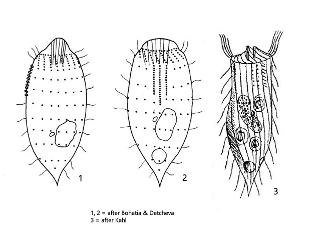
oral apparatus apical, cytopharynx with extrusomes
body uniformly cilated apart from the dome-shaped anterior end
a ring of elongated cilia borders the dome-shaped anterior end
macronucleus oval to kidney-shaped
one spherical micronucleus
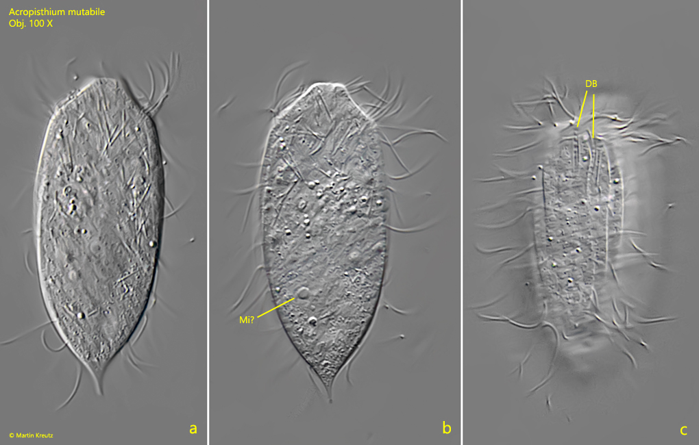
a dorsal brush consisting of three rows, one row with bristles, two rows with humps
contractile vacuole sub-terminal
Acropisthium mutabile
Acropisthium mutabile is described by Kahl as a very common species. I could find Acropisthium mutabile only a few times in Ulmisried and Simmelried. It is remarkable that I found the specimens exclusively in the cold season between end of October and mid of March. I could not find them in the summer months.
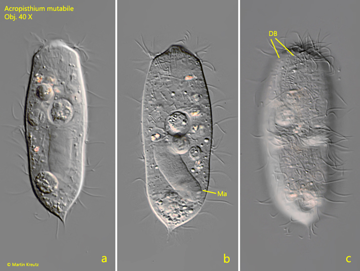
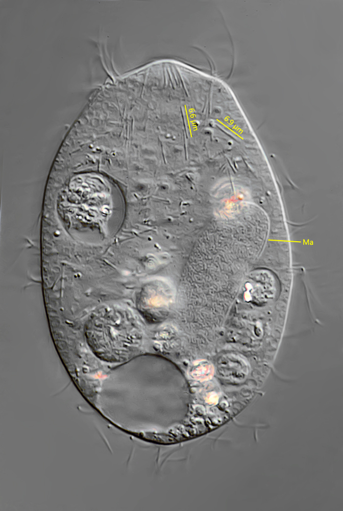
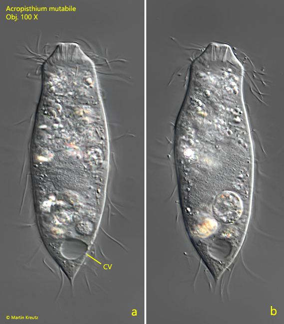
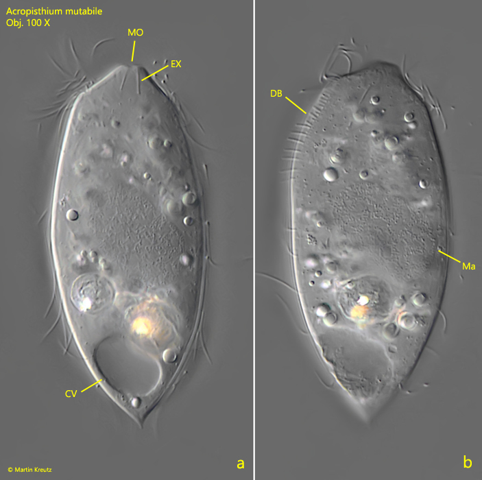
Although Acropisthium mutabile is mostly smaller than 100 µm, the species can be easily identified by the dome-shaped to snout-shaped anterior end and the pointed posterior end. The mouth opening is lined with rod-shaped, 6.6–6.9 µm long extrusomes (s. figs. 2 and 3a-b). The contractile vacuole is located sub-terminally (s. fig. 3a). In most specimens the macronucleus was long oval and slightly curved. I could recognize a dorsal brush comprising three rows and can confirm Kahl’s description that only one of the rows bears bristles, while the other two consist of small cusps or tubercles. According to Kahl, the diet should consist mainly of small algae. However, I could not find any phagocytosed algae in any of the specimens I examined. Although the contents of the food vacuoles are difficult to interpret, I consider their contents to be rather phagocytosed small ciliates or flagellates (s. figs. 2 and 3b).
Fig. 1 a-c:Acropisthium mutabile. L = 100 µm. A freely swimming specimen. DB = dorsal brush. Obj. 40 X.
Fig. 2:Acropisthium mutabile. The squashed specimen shown in fig. 1 a-c. Note the straight, rod-shaped extrusomes with a length of 6.6–6.9 µm. Ma = macronucleus. Obj. 100 X.
Fig. 3 a-b:Acropisthium mutabile. L = 72 µm. A freely swimming second specimen. Note the contractile vacuole (CV) located sub-terminally. Obj. 100 X.
Fig. 4 a-c:Acropisthium mutabile. L = 61 µm. A freely swimming third specimen. DB = dorsal brush. Obj. 40 X.
Fig. 5 a-b:Acropisthium mutabile. L = 67 µm. A freely swimming fourth specimen. Note the row of the dorsal brush (DB) with bristles (b). Obj. 100 X.