axiopodia thick, with granules, distally tapered, shorter than cell diameter
axonemes of the axiopodia orginate near the nuclei
Actinosphaerium eichornii
The specimens of Actinosphaerium eichhornii shown here are from the Mindelsee. However, I find the species frequently also in my other sampling areas, mostly in the uppermost leaf layer at the bottom.
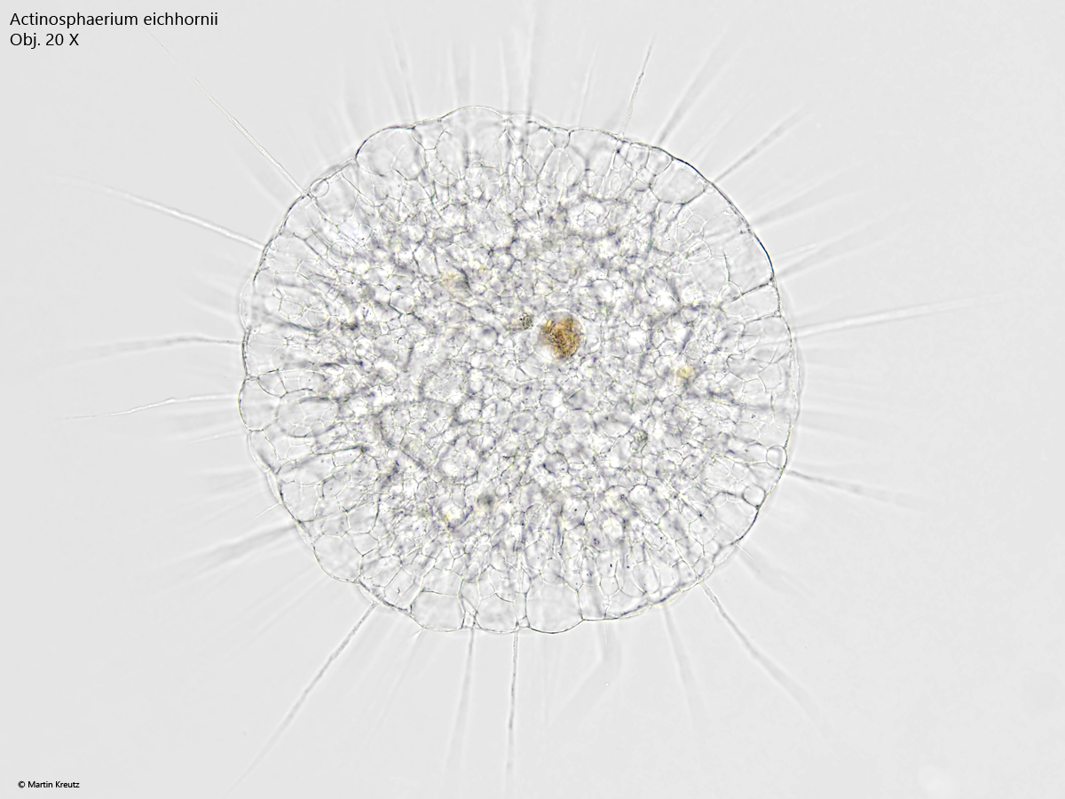
Often the specimens are non-transparent, due to phagocytozed prey (e.g. rotifers or ciliates). However, in my population, starving, very transparent specimens were also found, and their internal structure could be easily investigated (s. figs. 1 and 2).
Fig. 1: Actionosphaerium eichhornii. D = 450 µm. A slightly squashed specimen in brightfield illumination. The ectoplasm is the zone with large vacuoles while the central endoplasm consists of smaller vacuoles. Obj. 20 X.
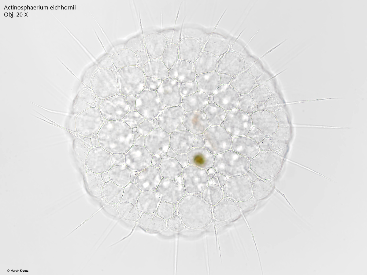
Fig. 2: Actionosphaerium eichhornii. D = 450 µm. A slightly squashed specimen in brightfield illumination. The focus is on the periphery of the ectoplasm. Obj. 20 X.
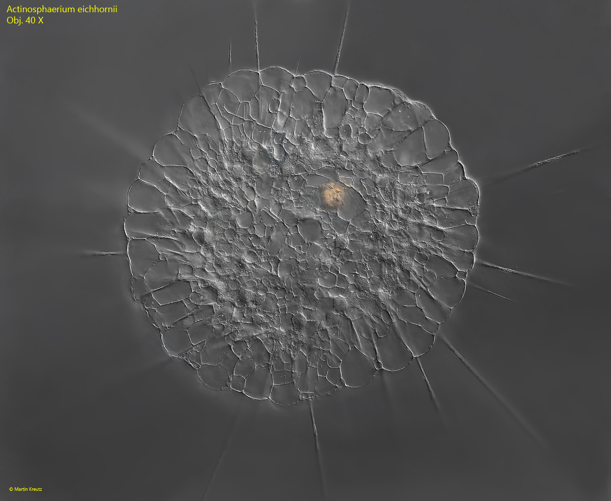
Fig. 3: Actionosphaerium eichhornii. D = 450 µm. The same, slightly squashed specimen as in figs. 1 and 2, but in DIC. The nuclei scattered in the endoplasm become visible. Obj. 40 X.
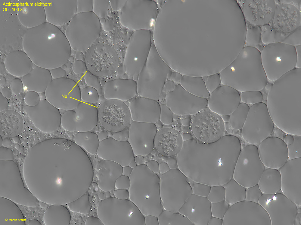
Fig. 4: Actionosphaerium eichhornii. Some of the nuclei (Nu) scattered in the endoplasm. Obj. 100 X.
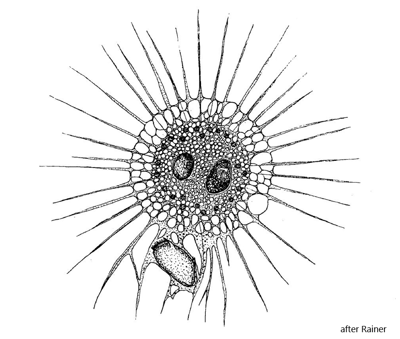
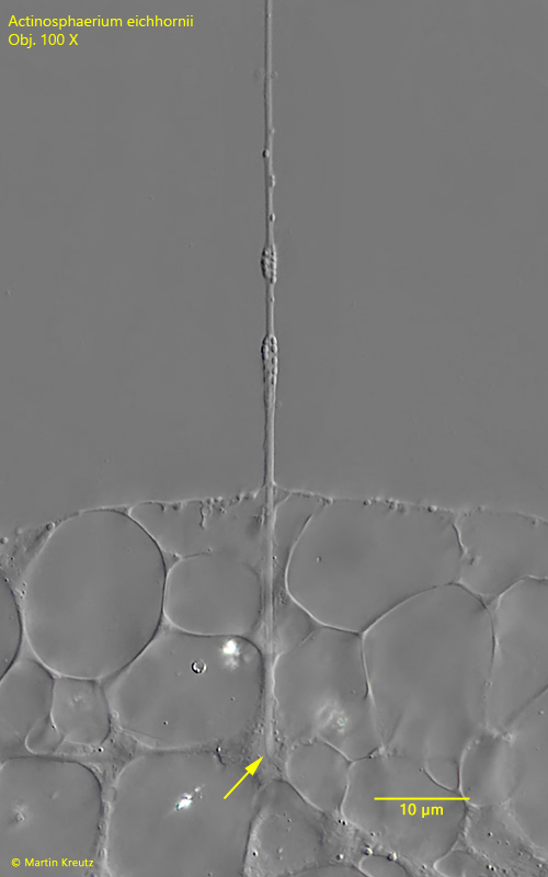
Actinosphaerium eichhornii belongs to the Actinophryidae, in which the microtubule bundles (axonemes) of the axopodia do not terminate in a centroplast (as in Centrohelids) but end freely in the plasm near the nuclei (s. fig. 5).
Fig. 5: Actionosphaerium eichhornii. D = 450 µm. The axoneme of the axiopodium terminate in endoplasm near a nucleus (arrow). Obj. 100 X.
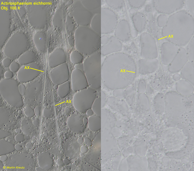
Fig. 6: Actinosphaerium eichhornii. D = 450 µm. Some axonemes (AX) of the axiopods in the ectoplasm of a strongly squashed specimen in DIC (left) and brightfield illumination (right). Obj. 100 X.
The axonemes are composed of bundles of microtubules. Under strong coverslip pressure, the microtubule bundles disintegrate into their individual microtubule fibers, which can be seen by light microscopy at the resolution limit (s. fig. 6).
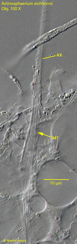
Fig. 7: Actinosphaerium eichhornii. Under strong coverslip pressure the axonemes disintegrate into the microtubule fibers (MT). Obj. 100 X.