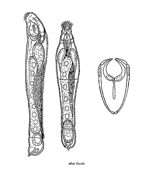
corona strongly reduced to semi-circular ciliary field
trophi small, unci needle-shaped
egg maturation in the posterior end of body
endoparasitic lifestyle in the intestine of Nais and Stylaria
Albertia naidis
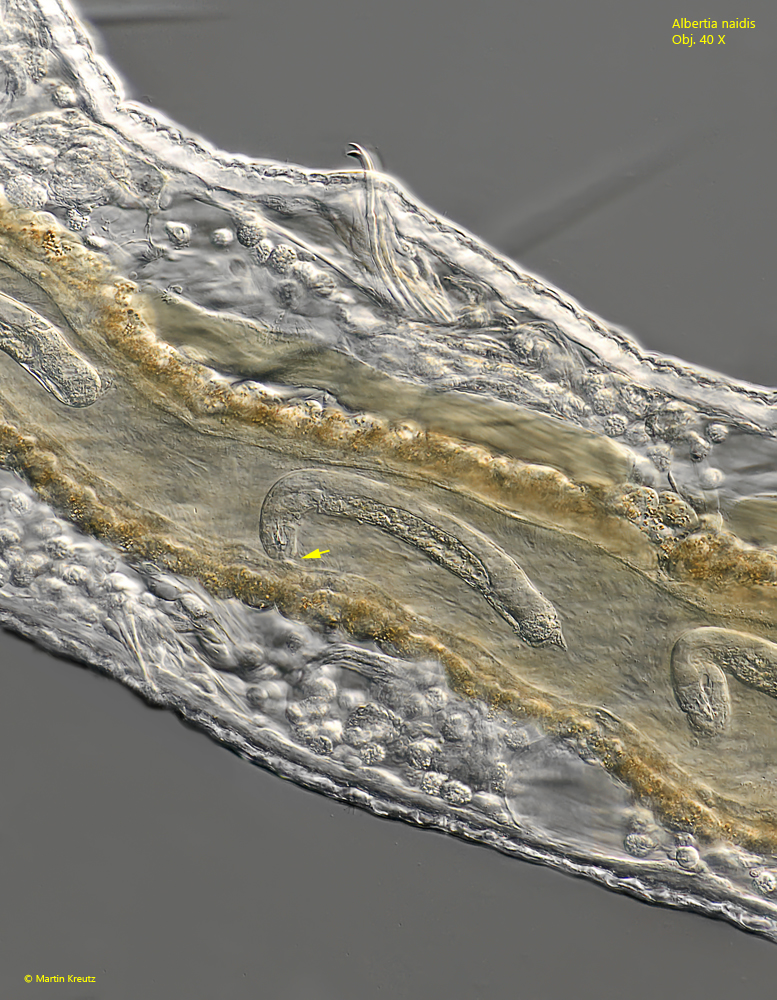
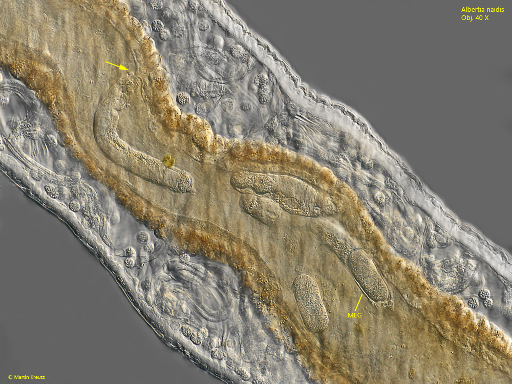
The rotifer Albertia naidis is an endoparasite living in the in the intestine of the Oligochaetes Nais spec. and Stylaria lacustris. With the pincer-like trophi Albertia naidis grasps the papillae of the epithelial cells in the intestinal wall to avoid being flushed out (s. figs. 2 and 4). In the lumen of the intestine, Albertia naidis feeds on the epithelial cells (Coineau & Kunst, 1964) and not on the ingested food of the Oligochaetes, which passes through the intestine.
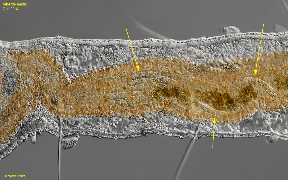
I have found Albertia naidis in old samples from the Simmelried, in which a high population of Nais spec. was present. In the free-moving Oligochaetes, Albertia naidis is difficult to detect because the intestine is often opaque due to ingested food and is also yellow-brown or red-brown in color due to oil droplets in the intestinal wall. Only in the slightly squashed specimens can the worm-like endoparasites be seen (s. fig. 1). In total I could find 4 specimens of Nais spec., which were infested by Albertia naidis. In their intestines 10 -20 specimens of the parasites were present each.
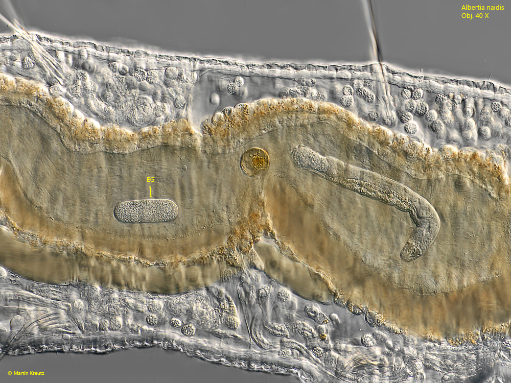
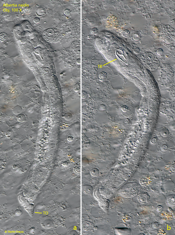
Albertial naidis forms quite large eggs, about 40–60 µm long, which mature in the posterior end and occupy almost one third of the body length (s. fig. 4). The eggs are deposited in the intestinal lumen and then flushed out with the intestinal content (s. fig. 3). Close examination of Albertia naidis is not possible until the specimens are flushed out of the intestine by strong pressure (s. fig. 5 a-b). Foot and toes are strongly degenerated. There is only one, conically shaped toe, which is shifted to the ventral side (s. fig. 6). The head is barely separated from the rest of the body. In my population, the specimens of Albertia naidis were 120–140 µm long, which is about in the middle of the range of 94–350 µm given by Koste (1978).
I did not observe Albertia naidis causing lasting damage to its host. Infested specimens of Nais spec. were agile and also not smaller than non-infested specimens.
Fig. 1:Albertia naidis. Some specimens in the intestine of Nais spec. (arrows). Obj. 20 X.
Fig. 2:Albertia naidis. L = 164 µm. A specimen is feeding on the intestinal epithelium of Nais spec. (arrow). Note the ventrally bent head what is a characteristic feature of this species. Obj. 40 X.
Fig. 3:Albertia naidis. L = 168 µm. Next to the specimen a deposited egg (EG) in the intestine of Nais spec. is visible. The egg is 54 µm long. Obj. 100 X.
Fig. 4:Albertia naidis. A specimen with a mature egg (MEG) in the posterior end of the body. Another specimen is sucking on the intestinal epithelium (arrow). Obj. 40 X.
Fig. 5 a-b:Albertia naidis. L = 136 µm. Two focal planes of a specimen flushed out. Note the smal, pincer-shaped trophi (TR). TO = toe. Obj. 100 X.
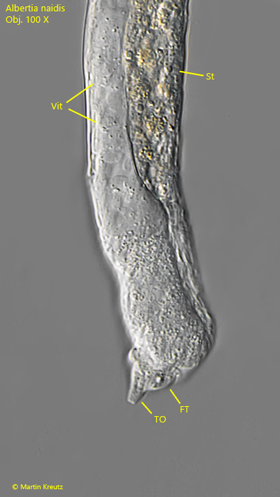
Fig. 6:Albertia naidis. The posterior end of a specimen in detail. Note the ventrally shifted toe (TO) and the strongly reduced foot (FT). Vit = vitellarium, St = stomach. Obj. 100 X.