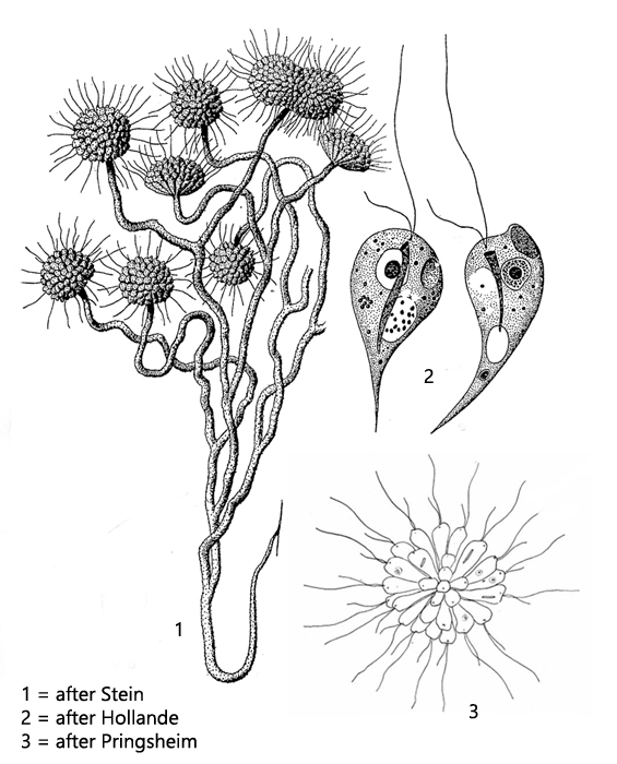
colorless flagellates with two flagella of different length
colonies on distal ends of branched, brownish stalks
stalks up to 1 mm long, covered with foreign material
average diameter of colony about 30 µm
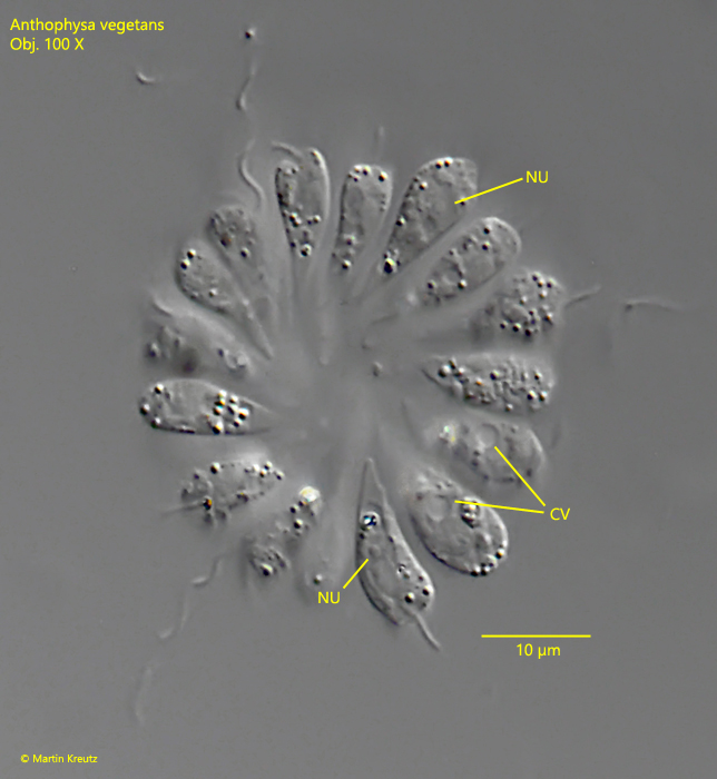
single cells 8 – 13 µm long
no eyespot
contractile vacuole in anterior third
spherical nucleus in mid-body
Anthophysa vegetans
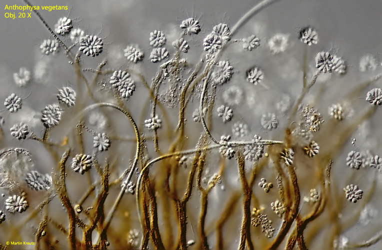
Colonies of Anthophysa vegetans can be often found in old samples on the surface or attached to the vessel wall, where they are easily recognized by their brownish coloration. Anthophysa vegetans is very common and I find this colorless flagellate in all my sampling locations. So the species seems to be very flexible in terms of living conditions. The colonies are constantly swirling bacteria for the flagellates to feed on. All populations I examined had the contractile vacuole located in the mid-body, which differs from the description by Stein, who located it in the anterior third of the body.
Fig. 1:Anthophysa vegetans. A large colony on branched, brownish stalks. Obj. 20 X.
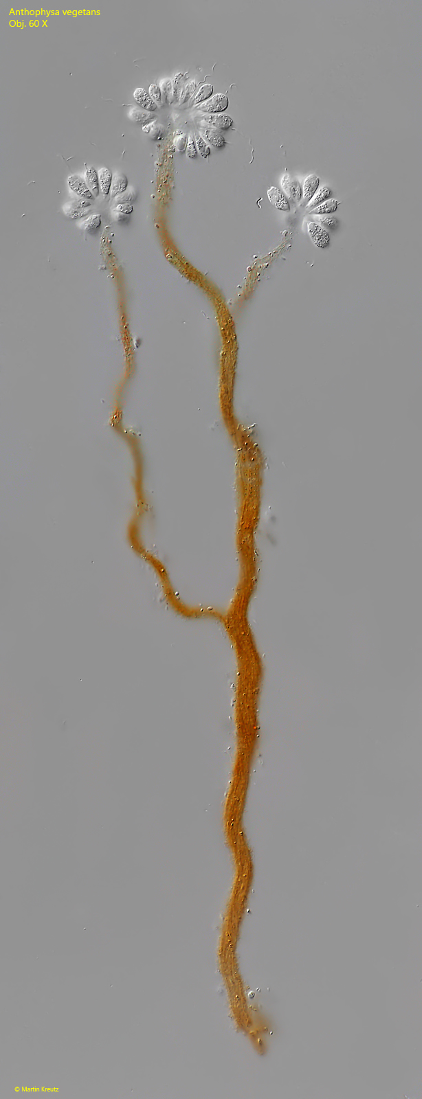
Fig. 2:Anthophysa vegetans. A small colony of about 60 individuals at the distal ends of the branched, brownish stalk. The length of the stalk with the colony is 480 µm. Obj. 100 X.
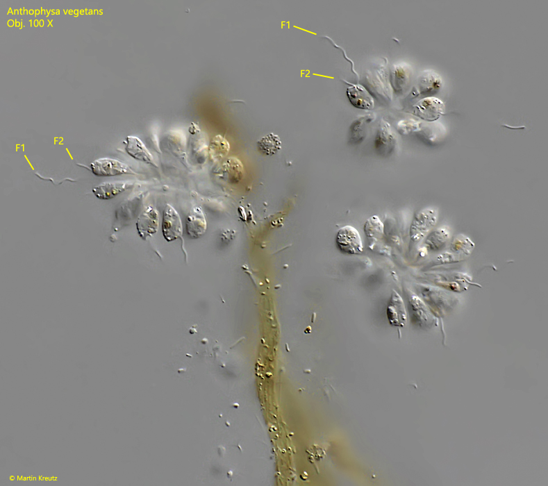
Fig. 3:Anthophysa vegetans. The colonies in detail. Each single cell has a long (F1) and a short flagellum (F2). Obj. 100 X.
Fig. 4:Anthophysa vegetans. L = 13 µm. Often detached colonies can be found. CV = contractile vacuole, NU = nucleus. Obj. 100 X.