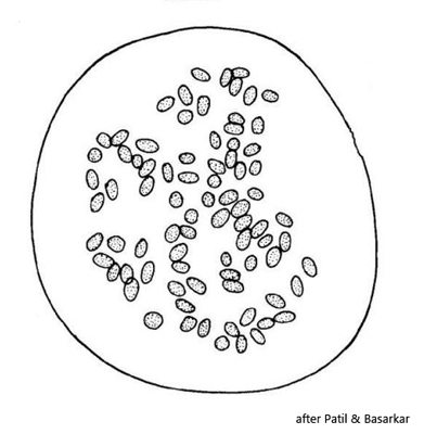
colony in a common mucilaginous sheath, irregularly or spherically shaped
cells loosely arranged
no individual mucilaginous envelope of cells
cells oblong, broadly rounded ends, sometimes slightly curved
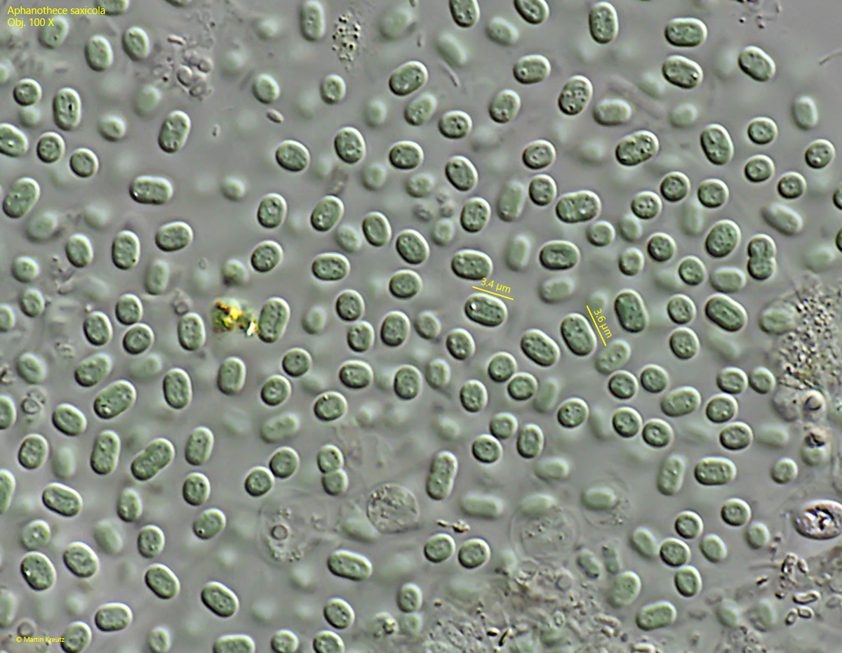
cells 1.7–2.6 µm broad, 3–6 µm long
color pale blue-green
cytoplasm homogenous, only few granules
gas vacuoles absent
Aphanothece saxicola
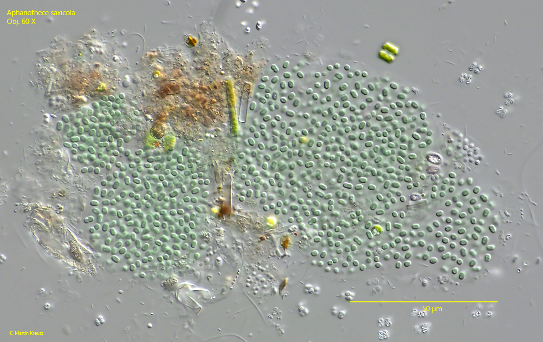
So far I have found Aphanothece saxicola in the Simmelried and in the pond of the convent of Hegne. There the colonies were found in the uppermost layer of mud.
The colonies were all between 70-100 µm in size and irregularly shaped. I did not find any spherical colonies. The distinction between the different species of the genus Aphanothece is essentially based on cell size, cell shape and habitat.
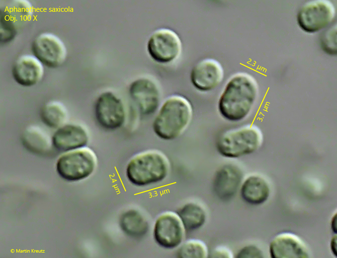
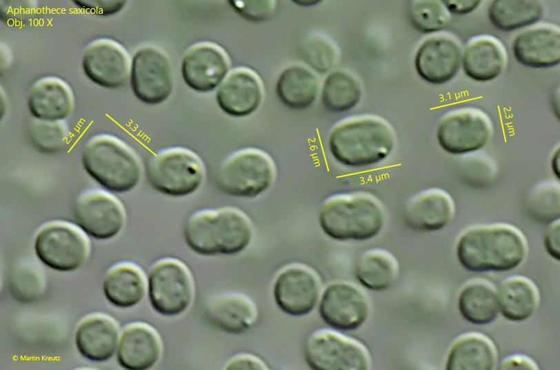
The cells in the colonies from both localities were very constant between 3.0–3.7 µm long and between 2.2–2.6 µm wide (s. figs. 2, 4 and 6). The cell shape was oblong, with broadly rounded ends. The similar species Aphanothece microscopica has larger cells (length = 3.2–10 µm, width = 3–6 µm).
Fig. 1:Aphanothece saxicola. L = 110 µm (of colony). A colony found in the Simmelried. Ob. 60 X.
Fig. 2:Aphanothece saxicola. L = 3.0–3.8 µm (of cells). The cells are broadly oblong. In the cytoplasm some granules or crystals are visible. Obj. 100 X.
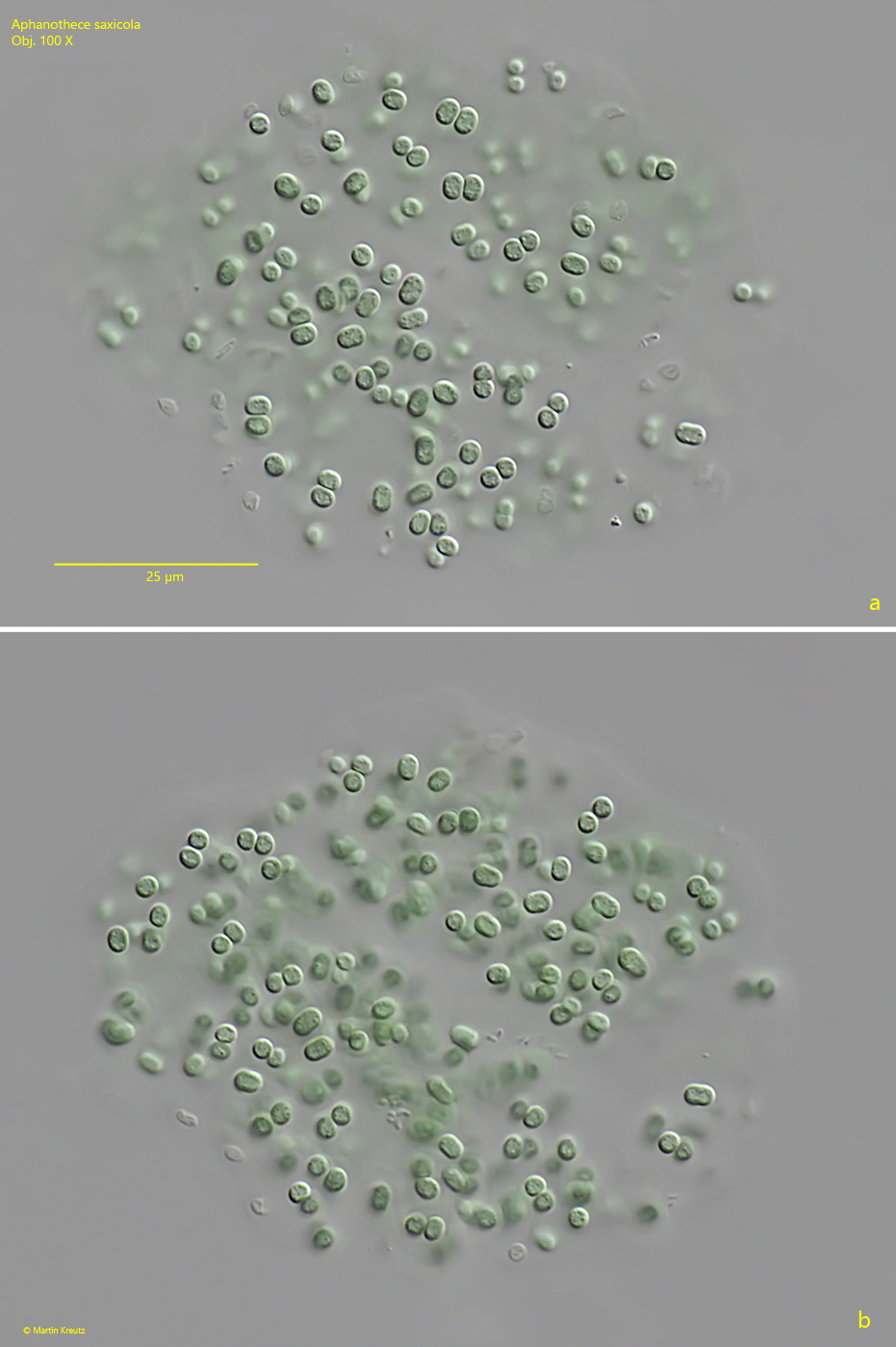
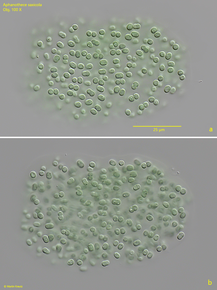
Fig. 3 a-b:Aphanothece saxicola. L = 70 µm (of colony). Two focal planes of a colony from the pond of the convent Hegne. Obj. 100 X.
Fig. 4:Aphanothece saxicola. L = 3.3–3.7 µm (of cells). Image section of fig. 3 a. Obj. 100 X.
Fig. 5 a-b:Aphanothece saxicola. L = 77 µm (of colony). Two focal planes of a second colony from the pond of the convent Hegne. Obj. 100 X.
Fig. 6:Aphanothece saxicola. L = 3.1–3.4 µm (of cells). Image section of fig. 5 a. Obj. 100 X.