basket of trichites straight to slightly oblique inserted
anterior end of basket with refractive collar
spherical macronucleus with central nucleolus or some peripheral nucleoli
macronucleus located in anterior half
one micronucleus adjacent to macronucleus
contractile vacuole in posterior third
about 30 µm long caudal cilium
extrusomes rod-shaped, 4 µm long
Apsiktrata gracilis
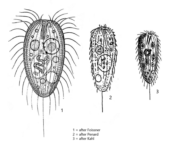
Apsiktrata gracilis is the most common prostomatid ciliate in the mud of Simmelried. It is present in almost all samples, sometimes in masses. The species can be easily identified by the thickened ends of the trichites of the basket, which together form an apikal, refractive collar (s. fig. 3). The basket of trichtites is often oblique relative to the longitudinal cell axis, which can be easily seen in freely swimming and spinning specimens. In addition, this ciliate has a rather long caudal cilium. In my population, all of the specimens I examined were filled with clearly visible symbiotic bacteria. Interestingly, these are not mentioned by any of the earlier authors, including Foissner, who published an accurate redescription in 1984 (still under Holophrya gracilis). Originally this species was described as Urotricha gracilis by Penard. Later Kahl placed it in Holophrya. Finally, Foissner, Berger and Kohmann transferred this species from the genus Holophrya to the newly created genus Apsiktrata, because in contrast to Holophrya and Prorodon species of the genus Apsiktrata do not possess a dorsal brush.
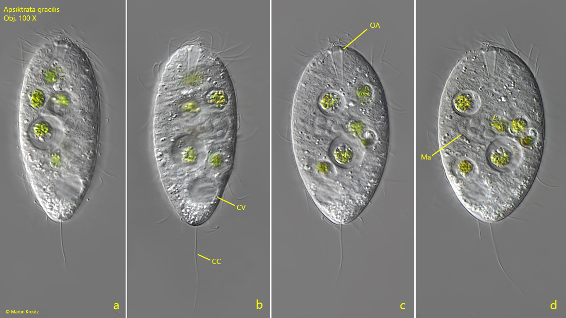
Fig. 1 a-d:Apsiktrata gracilis. L = 65 µm. A freely swimming specimen (a) is fixed and squashed during reduction of the layer thickness (b – c). CC = caudal cilium, CV = contractile vacuole, Ma = macronucleus, OA = oral aperture. Obj. 100 X.
Fig. 2 a-d:Apsiktrata gracilis. L = 52 µm. Different focal planes of a second, freely swimming specimen. Note the oblique basket of trichtites (BT). CC = caudal cilium, CV = contractile vacuole. Obj. 100 X.
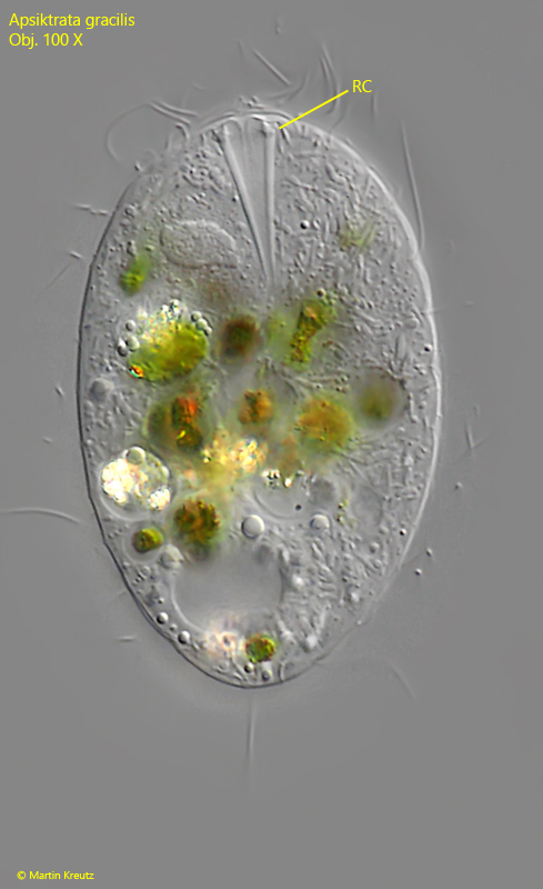
Fig. 3:Apsiktrata gracilis. L = 54 µm. This slightly squashed specimen has fed on small algae and cyanobacteria. Note the thickened anterior ends of the trichites appearing as a refractive collar (RC). Obj. 100 X.
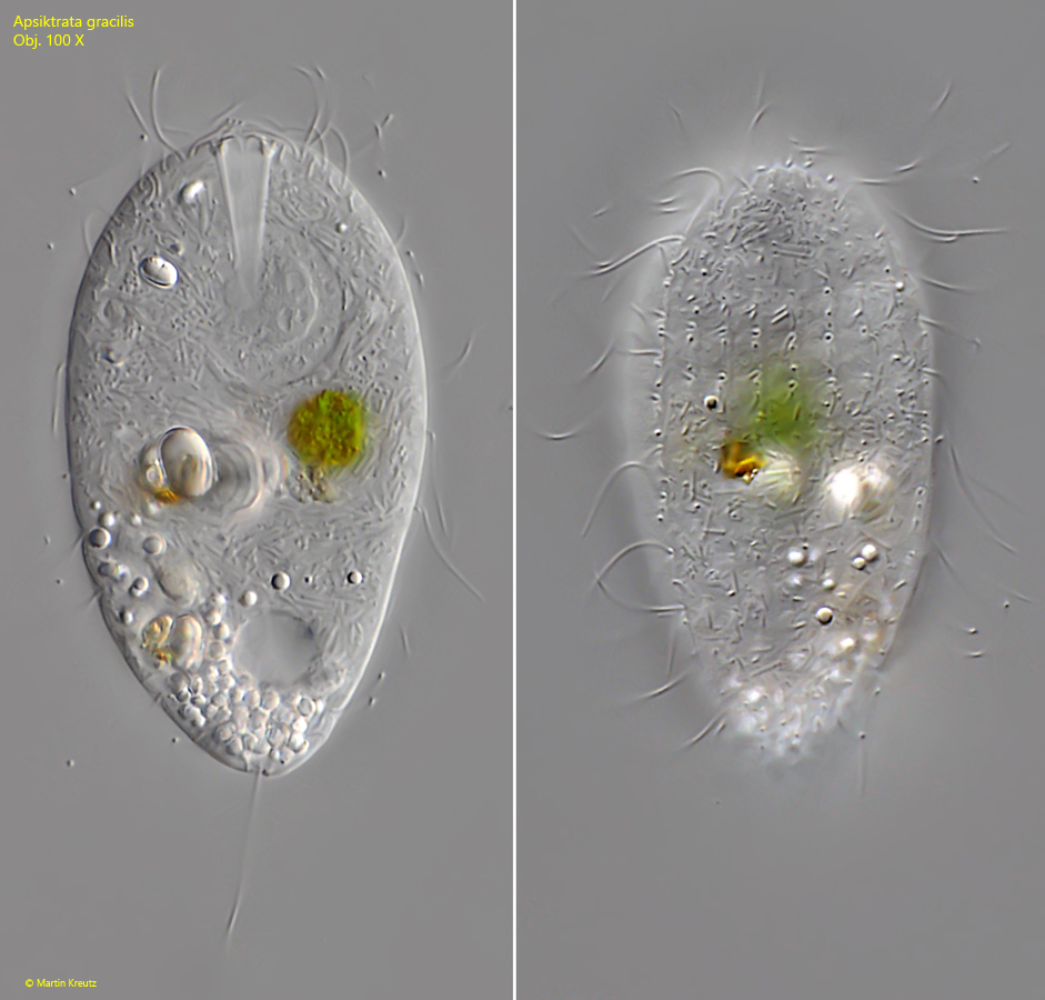
Fig. 4 a-b:Apsiktrata gracilis. L = 60 µm. Two focal planes of a slightly squashed specimen. Obj. 100 X.
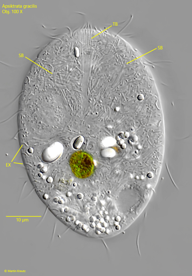
Fig. 5:Apsiktrata gracilis. The strongly squashed specimen shown in figs. 4 a-b. Beneath the pellicle the delicate extrusomes (EX) are visible. The cytoplasm is completely filled with symbiotic bacteria (SB) of different shape and size. TB = trichites of the basket. Obj. 100 X.
Fig. 6:Apsiktrata gracilis. L = 65 µm. Cyanobacteria and small green algae are often phagocytized as food. The digestion process in the food vacuoles sometimes results in colorful specimens. Ma = macronucleus, Mi = micronucleus. Obj. 100 X.