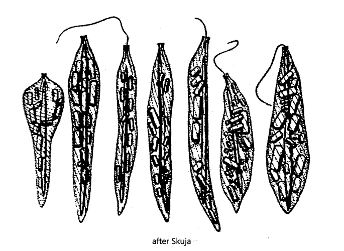
cell not very metoabolic, posterior and anterior end can be curved
delicate striation of the pellicle
one flagellum, about half body length
paramylon grains rod-shaped
a prominent, needle-shaped rod in the longitudinal axis of the cell, almost reaching body length
needle-shaped rod is birefringent and can break into parts
Astasia sagittifera
I find Astasia sagittifera exclusively in the Simmelried. Until about 15 years ago this species was quite common there, but since then it has become increasingly rare. The images of Astasia sagittifera shown below were taken in August 2006.
Astasia sagittifera is clearly set apart from similia species by a long, needle-like rod, which fills the entire length of the cell (s. figs. 1 a-b and 2 a). This limits the mobility of the cell. In addition to this prominent rod, other rod-shaped paramylon granules are found in the cell. The needle-shaped rod is not consisting of paramylon. This can be simply proved in polarized light, in which the needle-shaped rod shines brightly (i.e. is birefringent), while the paramylon grains remain dark.
The nature of the central rod was examined by Deflandre (1934). According to his investigations the substance is strongly birefringent, soluble in hydrocarbons such as gasoline, xylene, toluene, and chloroform but insoluble in ether and alcohols. It cannot be stained by osmic acid, Sudan III, iodine or ruthenium red. Up to now the chemical nature of the central rod could not be determined.
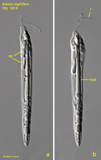
Fig. 1 a-b:Astasia sagittifera. L = 52 µm. A freely swimming specimen. Note the prominent needle-shaped rod (NSR) arranged in the longitudinal axis of the cell. F = flagellum, PB = rod-shaped paramylon bodies. Obj. 100 X.
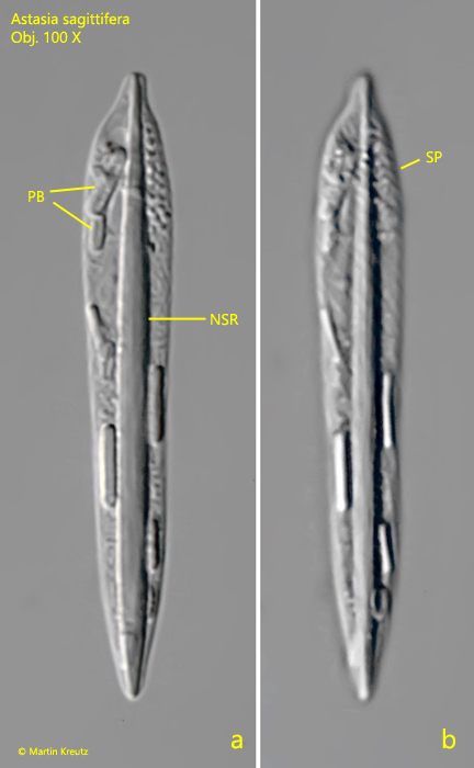
Fig. 2 a-b:Astasia sagittifera. L = 56 µm. Two focal planes of a strongly squashed specimen. The needle-shaped rod (NSR) is as long as the cell. PB = rod-shaped paramylon bodies, SP = striation of the pellicle. Obj. 100 X.
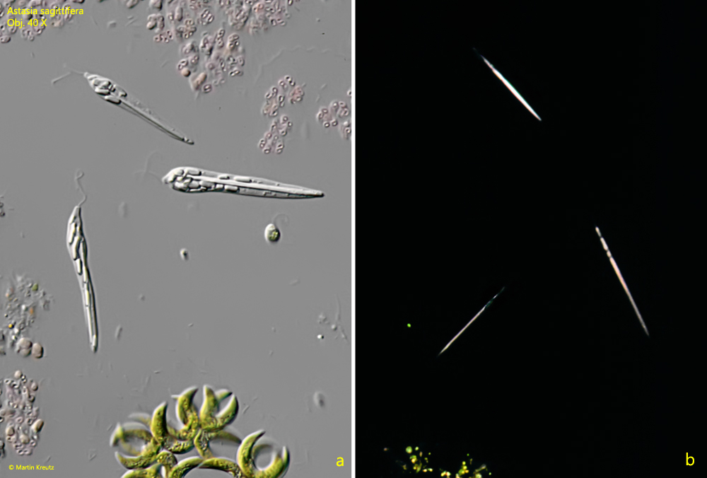
Fig. 3 a-b:Astasia sagittifera. L = 44 – 54 µm. Three freely swimming specimens in DIC (a) and in polarized light (b). The needle-shaped rods are birefringent and therefore shine brightly in polarized light. Obj. 100 X.