average length of cells 60–85 µm, average width 2–4 µm
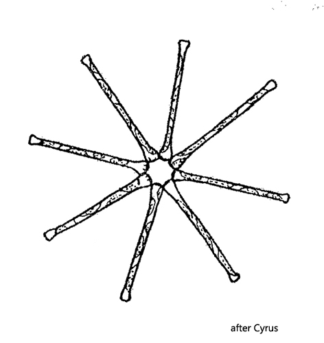
living cells joined in star-shaped colonies consisting of 8–20 cells
cells in colonies connected with a jelly at the apices
apices in the center of the colonies more strongly swollen than distal apices
6–8 golden-brown chromatophores per cell
nucleus in center of cell
Asterionella formosa
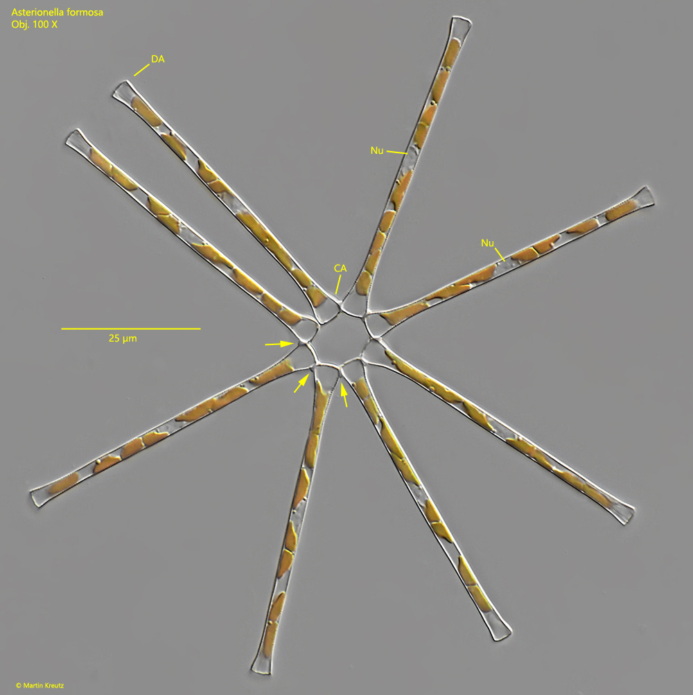
Asterionella formosa is one of the most common diatoms in the phytoplankton of water bodies. In Lake Constance it occurs partly in masses. The star-shaped colonies are easy to recognize. Often, however, the colonies are incomplete or have a spirally structure. Completely closed rings as shown in figs. 1 and 2 are more rarely found. The colonies are often colonized by small, sessile choanoflagellates. Colonies infested by a parasitic chytrid fungus (Zygorhizidium planktonicum) are also found. Infected cells often undergo apoptosis to stop a further spread of the fungus.
Fig. 1:Asterionella formosa. L = 56–58 µm (of the cells). A slightly squashed colony consisting of 8 cells. The cells are connected via the central apices (CA) with jelly (arrows). Note, that the central apices (CA) are more strongly swollen than the distal apices (DA). The nucleus (Nu) is located in the center of the cells. Obj. 100 X.
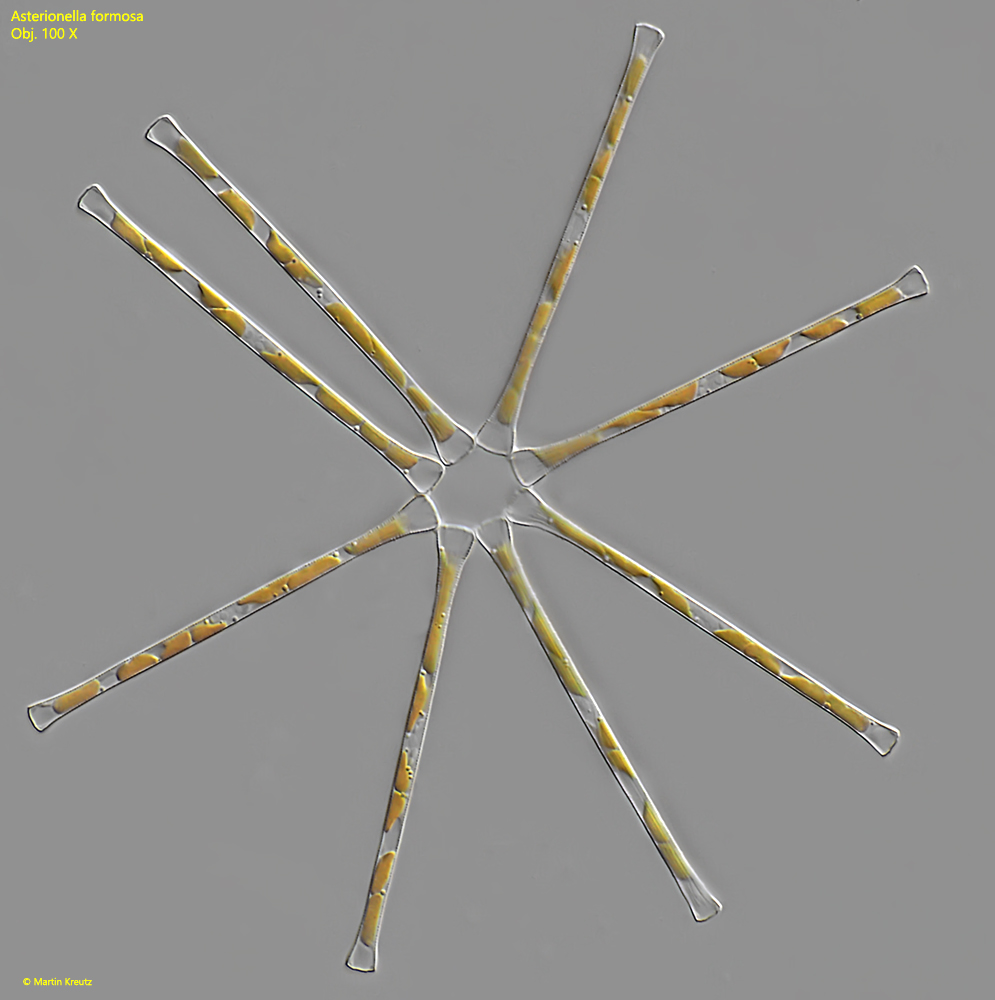
Fig. 2:Asterionella formosa. L = 56–58 µm (of the cells). The same colony as shown in fig. 1 but with a slightly different focal plane. Obj. 100 X.