body discoid, laterally compressed with flat left and convex right side
one ventral spine (above the adoral zone of membranelles)
four posterior spines
anterior extension of the dorsal keel in a teeth-like spine
short perizonal row of cilia
short terminal row of cilia on left side
one spherical macronucleus with one adjacent micronucleus
contractile vacuole in posterior third
Atopodinium fibulatum
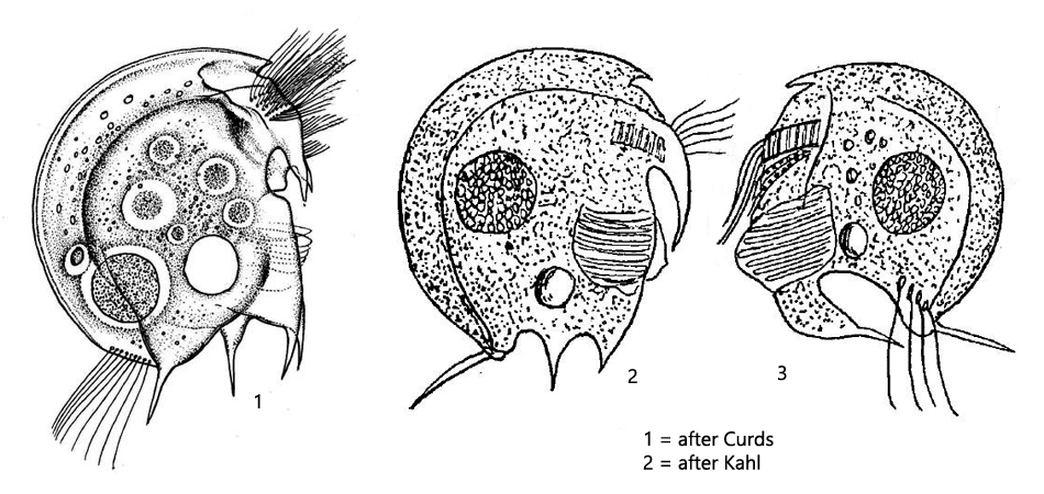
I have found Atopodinium fibulatum in all my sites where there is a mud layer of decomposing leaves and plant material. The species is very common and I find it throughout the year. This odontostomatid ciliate has reduced its somatic cilia to an extreme dregree. There is only a short row of cilia on the left side at the posterior end (hard to see) and the perizonal row above the adoral zone. The food is mainly guided by the perizonal cilia band, which is why the swimming style is very slow and jerky. Very characteristic in this species is a ventral spine over the adoral zone and a terminal spine pointing to the dorsal side. In my population the dimensions of the spines corresponded more to the drawing of Curds (s. drawing 1), while Kahl drew them much larger and more prominent (s. drawing 2 and 3). On the the hand, Kahl drew the frontal tooth much more pointed and smaller. This shows that the odontostomatid ciliates can be quite variable, which I have also noticed in other species and which makes correct identifications more difficult.
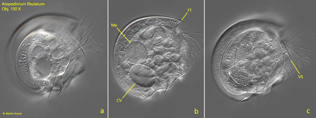
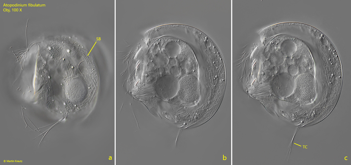
Fig. 1 a-c:Atopodinium fibulatum. L = 60 µm. Three focal planes of a slightly squashed specimen from the right side. Note the characteristic ventral spine (VS). CV = contractile vacuole, FT = frontal teeth, Ma = macronucleus. Obj. 100 X.
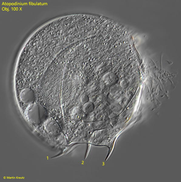
Fig. 2:Atopodinium fibulatum. L = 60 µm. The strongly squashed specimen shown in fig. 1 a-c for better visualisation of the posterior spines 1–3. Note that the spine 1 points dorsally in a characteristic way. Obj. 100 X.
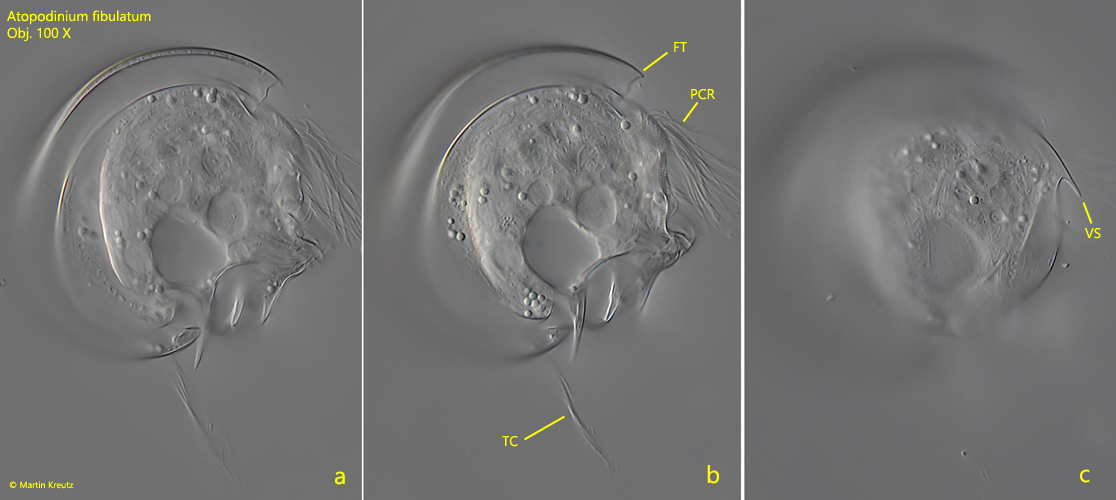
Fig. 3 a-c:Atopodinium fibulatum. L = 56 µm. Three focal planes of a second specimen from the right side. Note the tuft of terminal cilia (TC) arising from a short row on the left side of the body. FT = frontal teeth, PCR = perizonal row of cilia, VS = ventral spine. Obj. 100 X.
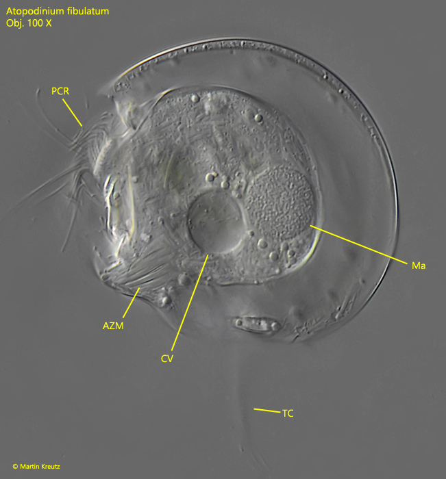
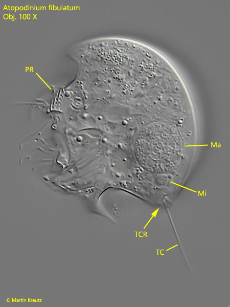
Under the coverslip, Atopodinium fibulatum is most often found lying on the left side with the right side facing the coverslip. This is probably because the left side is flat while the right side is convex. This is why it is rare to find specimens lying on the right side to investigate the left side (s. figs. 4, 5 and 6 a-c).
Fig. 4:Atopodinium fibulatum. L = 53 µm. A slightly squashed specimen from the left side. AZM = adoral zone of membranelles, CV = contractile vacuole, Ma = macronucleus, PCR = perizonal row of cilia, TC = terminal cilia. Obj. 100 X.
Fig. 5:Atopodinium fibulatum. L = 40 µm. A second specimen from the left side. Note the short row of terminal cilia (TCR). Only one of the cilia is in focus (TC). Ma = macronucleus, Mi = micronucleus, PR = perizonal row. Obj. 100 X.
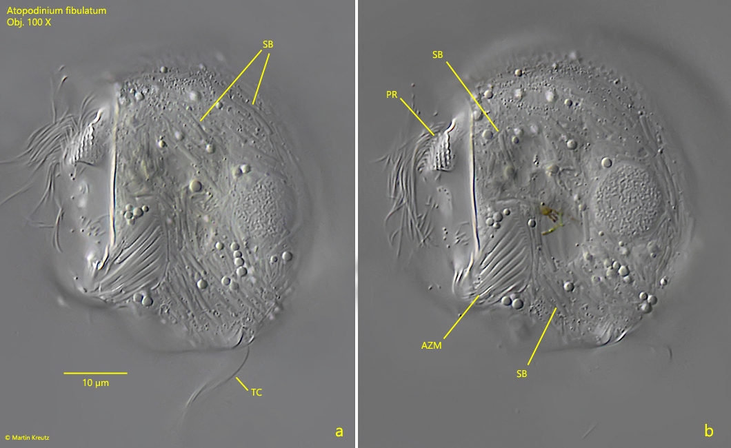
Fig. 6 a-c:Atopodinium fibulatum. L = 54 µm. A slightly squashed specimen from the left side. SB = symbiotic bacteria, TC = terminal cilia. Obj. 100 X.
In my population of Atopodinium fibulatum I have found many specimens which are completely filled with symbiotic bacteria (s. fig. 6), while in other specimens only very few symbiotic bacteria were visible or none at all. The reason for this variability is not known to me. Possibly it depends on the environmental conditions, i.e. in which depth the specimens are in the mud. The symbiotic bacteria are rod-shaped and with 5–8 µm length quite large and therefore clearly visible (s. fig. 7 a-b).
Fig. 7 a-b:Atopodinium fibulatum. Two focal planes of a slightly squashed specimen from the left side filled with symbiotic bacteria (SB). The bacteria are rods with a length of 5 – 8 µm. AZM = adoral zone of membranelles, PR = perizonal row, TC = terminal cilia. Obj. 100 X.