adoral zone starts on dorsal side and runs obliquely to the ventral side
perizonal stripe runs in parallel to adoral zone
mouth opening below equatorial zone
posterior end obliquely truncate
pellicle with distinct longitudinal ribs
contractile vacuole terminal
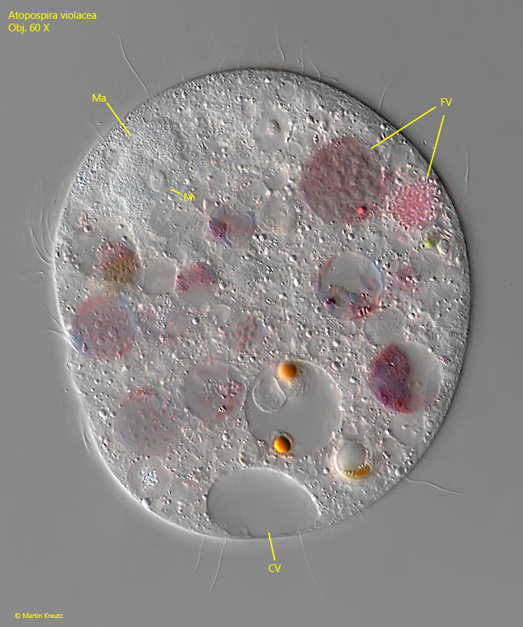
macronucleus globular or oval with adjacent spherical micronucleus
cytoplasm filled with ingested colonies of rhodobacteria
posterior end with some caudal cilia
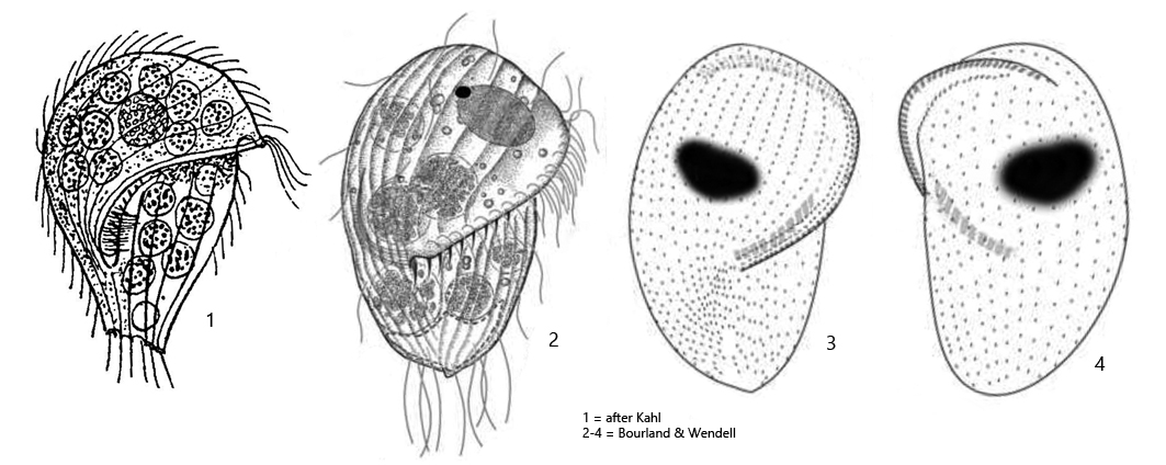
Atopospira violacea
Atopospira violacea was first described in 1926 by Kahl as Metopus violaceus. In 2014 the species was redescribed by Bourland & Wendell who transferred the species to the genus Atopospira. The genus Atopospira was erected by Jankowski in 1964.
So far I could detect Atopospira violacea only in Ulmisried and Simmelried. The ciliate is easy to recognize, because it is always purple colored due to the many food vacuoles, which are filled with ingested rhodobacteria. Mostly it becomes longer than 100 µm and is therefore quite stately. The distinctly ribbed pellicle is also conspicuous, which is especially noticeable on the dorsal side (s. fig. 3). Atopospira violacea swims very slowly and is easy to track. The adoral zone has only a few membranelles (about 10) and starts on the dorsal side at the anterior end (s. fig. 3). From there it runs obliquely across the ventral side to just below the cell equator (s. fig. 2 a-b). Due to the many food vacuoles, the cell content is not easy to investigate. The globular macronucleus and the adjacent micronucleus are only well recognizable in squashed specimens (s. fig. 4). Atopospira violacea has elongated caudal cilia, but they are very delicate (s. fig. 1 d) and not easy to recognize.
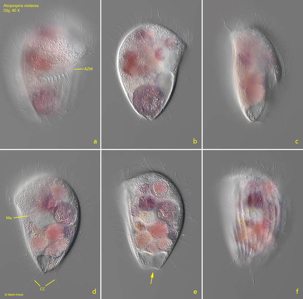
Fig. 1 a-f:Atopospira violacea. L = 104 µm. A freely swimming specimen from ventral (a, b), right (c) and from dorsal (d, e. f). Note the truncated posterior end, sometimes with a concave depression (arrow) depending on the filling level of the contractile vacuole. AZM = adoral zone of membranelles, CC = caudal cilia, Ma = macronucleus. Obj. 40 X.
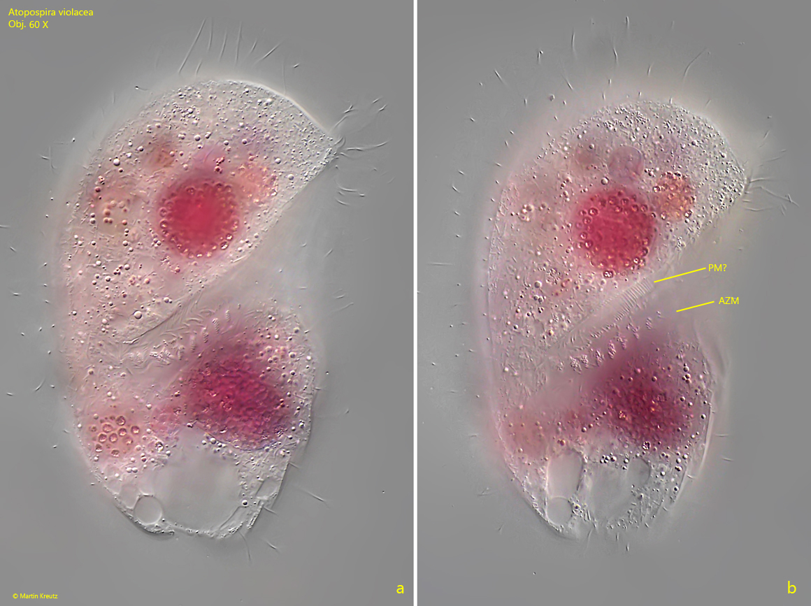
Fig. 2 a-b:Atopospira violacea. L = 133 µm. Two focal planes of a slightly squashed specimen from ventral. AZM = adoral zone of membranelles, PM? = likely the paroral membrane. Obj. 60 X.
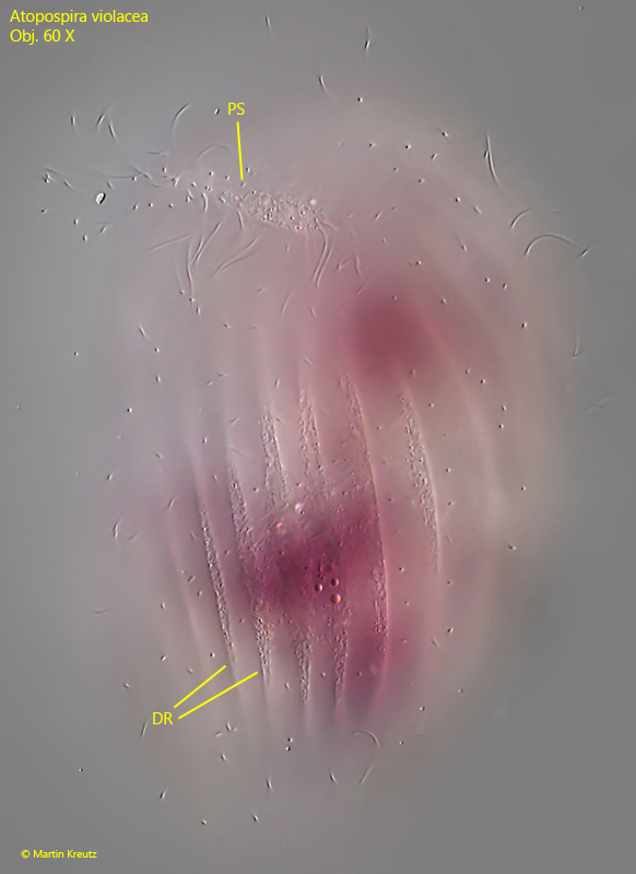
Fig. 3:Atopospira violacea. L = 122 µm. Focal plane of the dorsal side with the distinct ribs (DR) of the pellicle. Anteriorly the dorsal part of the perizonal stripe (PC) is visible. Obj. 60 X.
Fig. 4:Atopospira violacea. A strongly squashed specimen. CV = contractile vacuole, FV = food vacuoles, Ma = macronucleus, Mi = micronucleus. Obj. 60 X.