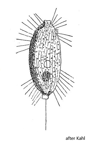
with short collar-like projections at anterior and posterior ends
contractile vacuole almost central
spherical macronucleus in posterior third
oral opening similar to Loxocephalus (hard to see)
gap of ciliation in mid-body
one long caudal cilium
Balanonema biceps
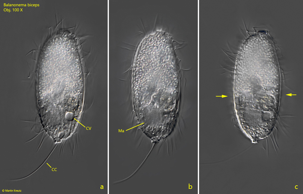
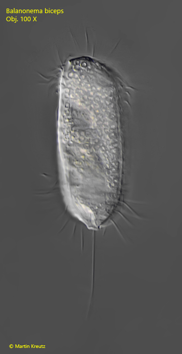
During my investigations of the Simmelried since 1993 I found only two specimens of Balanonema biceps in May 2008 (s. fig. 1 a-c) and in October 2008 (s. fig. 2).
The species was first described by Penard as Uronema biceps (1922). Kahl then later placed it in Balanonema. There are two further, similar species in the genus Balanonema. The first is Balanonema dubium (Penard, 1922) Kahl 1931, which however has flatter projections on the anterior and posterior ends and is also ciliated throughout. The other species is Balanonema sapropelica (Foissner, 1978), which has similarly shaped projections as Balanonema biceps, but is also ciliated in the middle of the body. For the differentiation of the three species both the shape of the projections and the ciliation of the body is important.
Deviating from the description and drawing of Kahl (s. drawing above), the contractile vacuole of my specimen is localized in the posterior third (s. fig. 1a). Since I have found only two specimens so far, I cannot judge if this is within the variability of the species.
Fig. 1 a-c: Balanonema biceps. L = 46 µm. Three focal planes of a slightly squashed specimen. Note the gap of ciliation in mid-body (arrows). CC = caudal cilium, CV = contractile vacuole, Ma = macronucleus. Obj. 100 X.
Fig. 2: Balanonema biceps. L = 50 µm. A second specimen found in October 2008. Obj. 100 X.