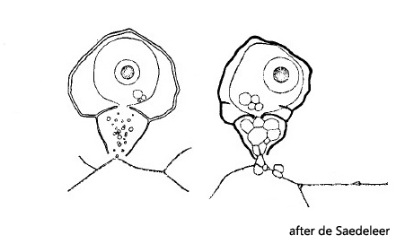
shell divided into two chambers, separated by a diaphragm with a porus
porus in the diaphragm often tube-shaped
nucleus central with a large sphaerical nucleolus
one contractile vacuole
shell often covered with ironoxide deposits
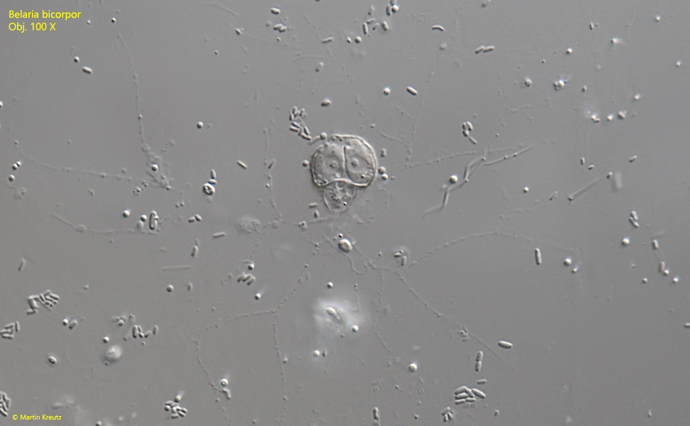
granuloreticulopodia forming a widely spreaded reticulum
Belaria bicorpor
I find the granuloreticulose testate amoeba Belaria bicorpor exclusively in the Simmelried. However, it is practically never found in the samples because it is often attached to detritus particles and then cannot be detected because of its small size. A simple method to extract it from the samples is the “floating coverslip”. You put some sample in a petri dish and put some coverslips on the water surface (they float). After 2–3 days many species have already settled and among them I regularly find Belaria bicorpor. Since the amoebae build their shells directly on the coverslip, they can then be easily observed at high magnifications.
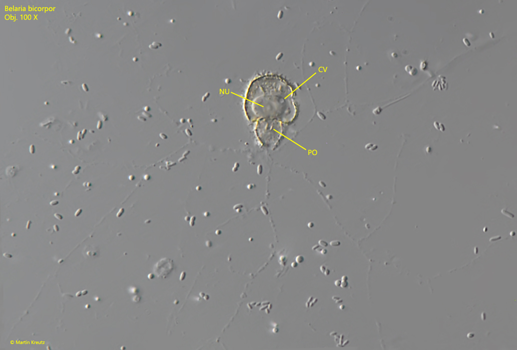
Fig. 1: Belaria bicolor. L = 15 µm. A specimen attached to the coverslip with a widely spreaded reticulum of granuloreticulopodia. Note the tube shaped porus (PO) in the diaphragm separating the two chambers of the shell. CV = contractile vacuole, NU = nucleus. Obj. 100 X.
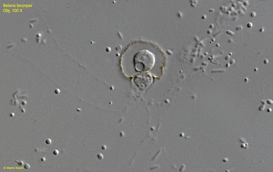
Fig. 2: Belaria bicolor. L = 13 µm. A young specimen with a thin shell. Obviously the second chamber is just under construction and filled with cytoplasm. Obj. 100 X.
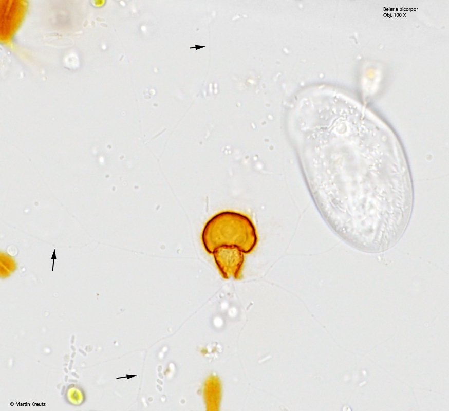
Fig. 3: Belaria bicolor. L = 14 µm. A specimen with a widely spreaded reticulum of granuloreticulopodia in brightfield illumination. Note the delicate granuloreticulopodia (arrows) and the orange-brownish color of the shell. Obj. 100 X.
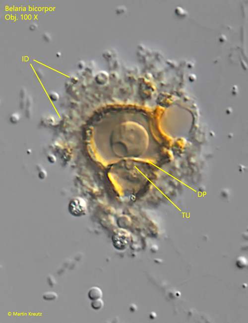
Fig. 4: Belaria bicolor. L = 14 µm. A specimen covered with deposits of ironoxide (ID). DP = diaphragm, TU = tube-shaped porus. Obj. 100 X.
Fig. 5: Belaria bicolor. L = 13 µm. A specimen after cell division. One of the specimens will leave the shell to construct its own. Obj. 100 X.