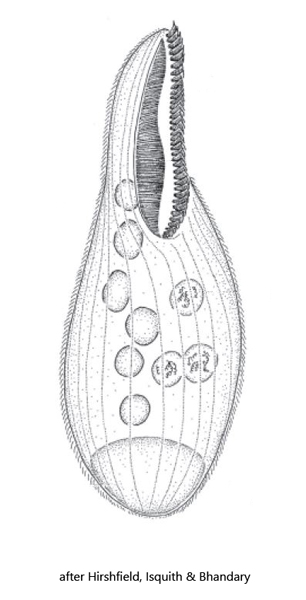
body long oval, laterally flattened, non-contractile
length 180–280 µm
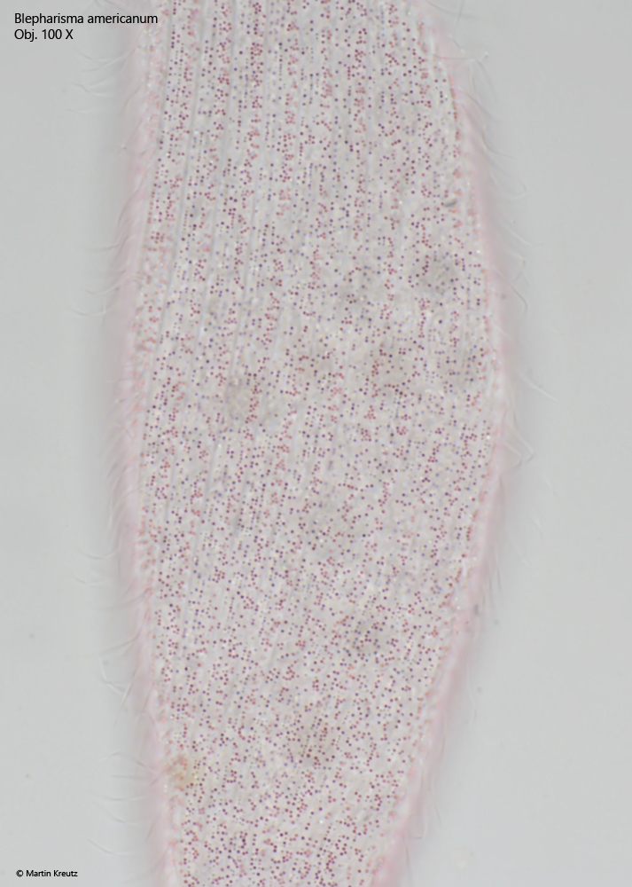
pigmentation pink
pigmentation granules in longitudinal bands
mouth opening at two fifths of the body length or mid-body
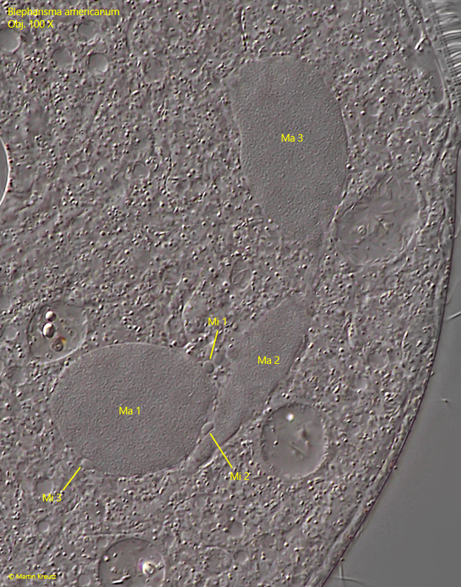
moniliform macronucleus with 3–8 nodules
6–20 small micronuclei adjacent to the nodules of the macronucleus
contractile vacuole terminal
Blepharisma americanum
Blepharisma americanum can be distinguished from the similar species Blepharisma undulans by the nuclear apparatus. While Blepharisma americanum has a moniliform macronucleus with 3-8 nuclear nodules, Blepharisma undulans has a binodal macronucleus, which consists of two nuclear parts connected by a filament. Since I was able to detect more than 2 nuclear nodules, Blepharisma americanum must be present here. I find Blepharisma americanum rarely in the Simmelried, mostly between decomposing plant masses. The intense purple color makes this large ciliate very photogenic.
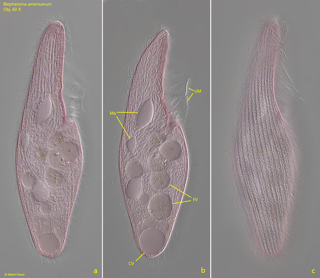
Fig. 1 a-c:Blepharisma americanum. L = 190 µm. Three focal planes of a freely swimming specimen. CV = contractile vacuole, FV = food vacuoles, Ma = nodules of the moniliform macronucleus, UM = undulating menbrane. Obj. 60 X.
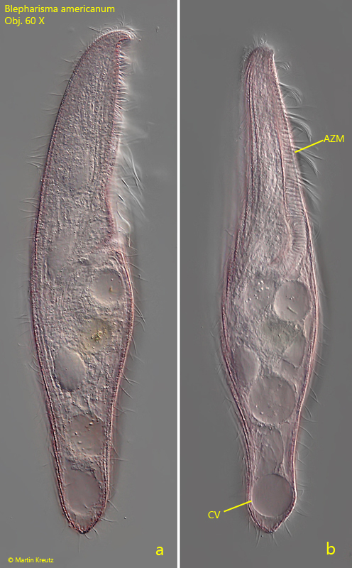
Fig. 2 a-b:Blepharisma americanum. L = 240 µm. A second freely swimming specimen from right (a) and from ventral (b). AZM = adorale zone of membranelles, CV = contractile vacuole. Obj. 60 X.
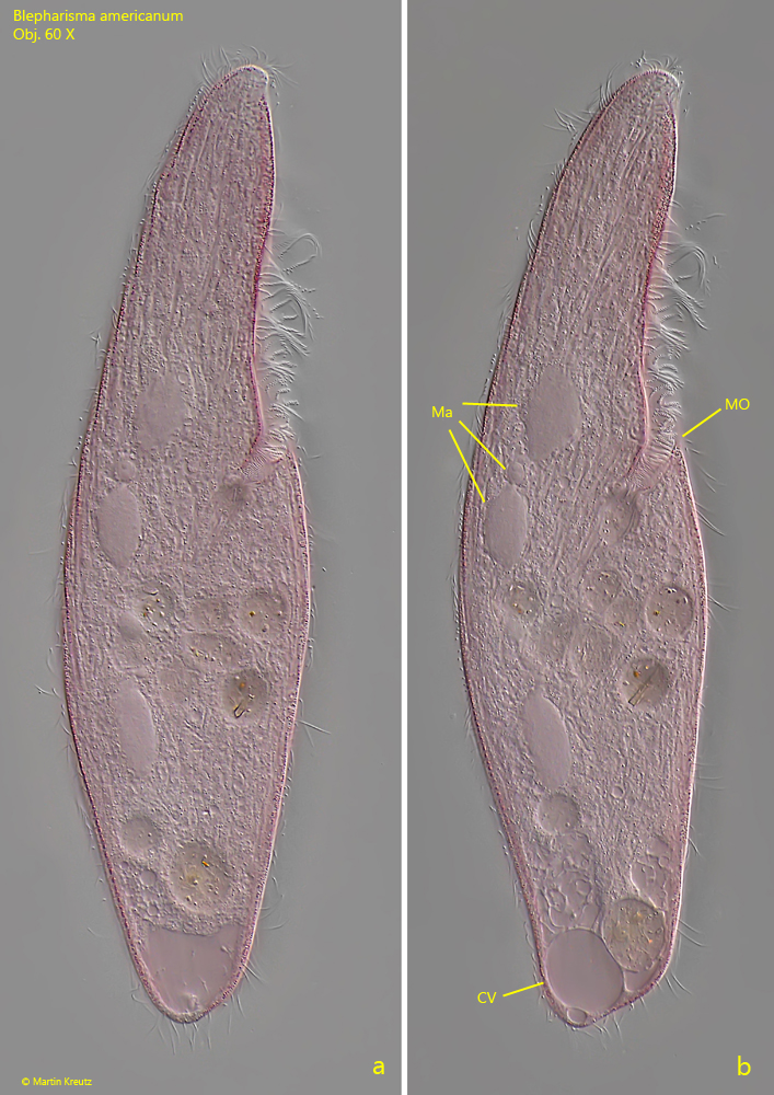
Fig. 3 a-b:Blepharisma americanum. L = 262 µm. A third, slightly squashed specimen. Ma = nodules of the moniliform macronucleus, MO = mouth opening. Obj. 100 X.
Fig. 4:Blepharisma americanum. The micronuclei (Mi 1–3) are adjacent to the nodules of the moniliform macronucleus (Ma 1–3). Obj. 100 X.
Fig. 5:Blepharisma americanum. The pink colored granules arranged in the pellicle in brightfield illumination. Obj. 100 X.