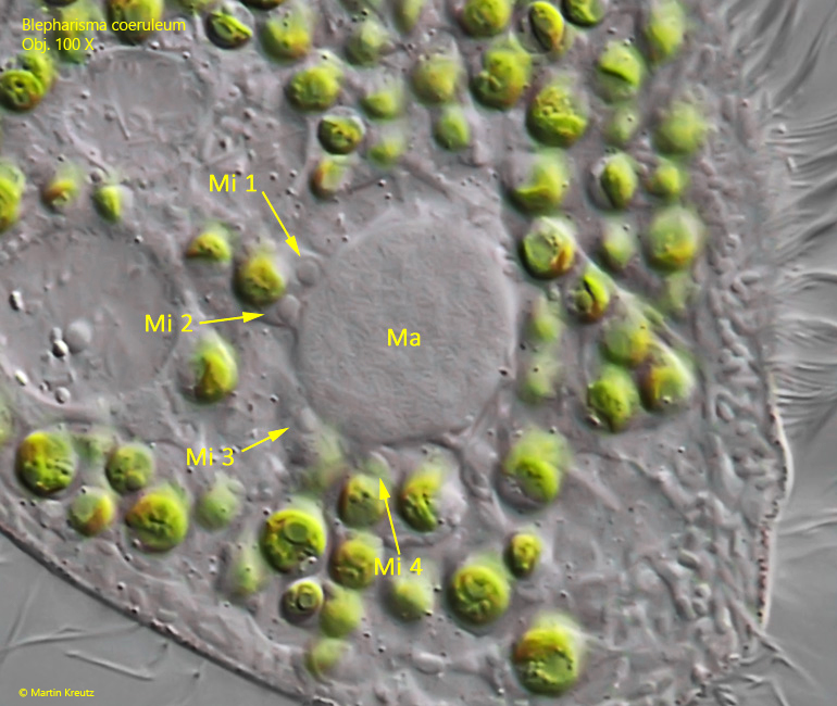
macronucleus spherical to oval with 3–6 adjacent micronuclei
granula bluish or green-bluish
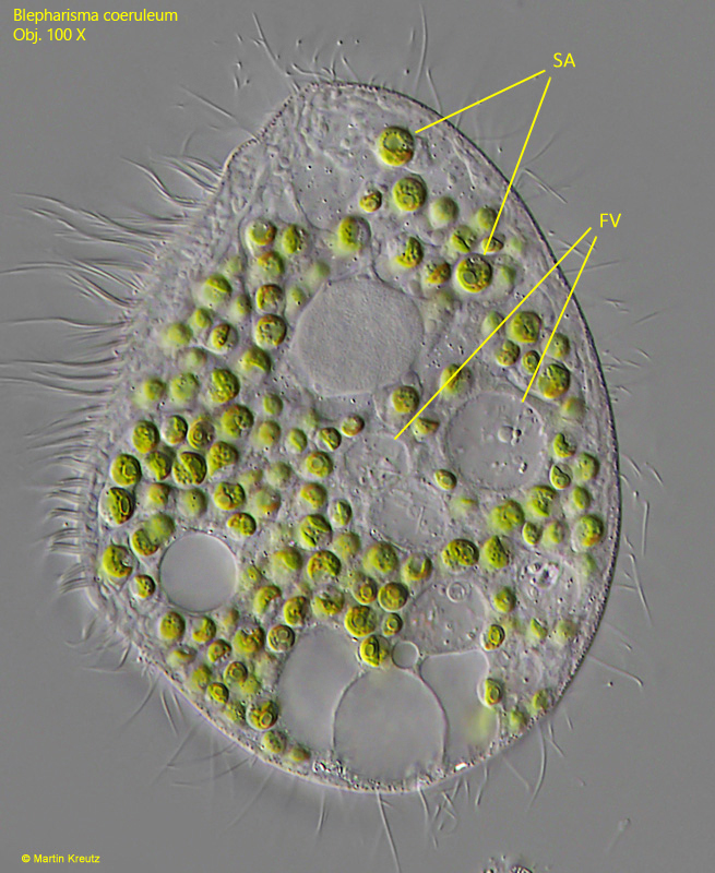
contains symbiotic algae
adorale zone reaches third of body
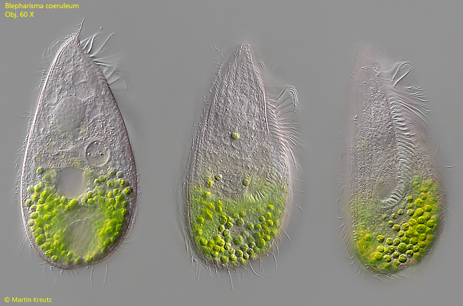
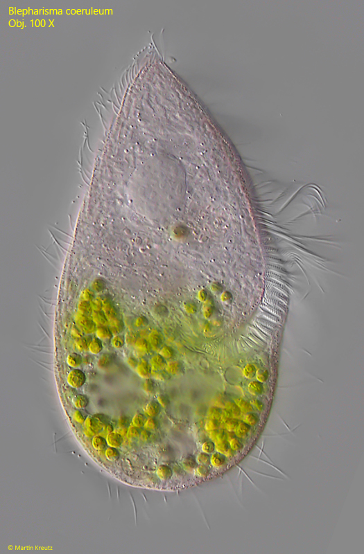
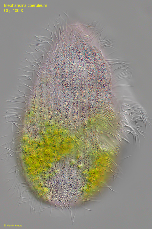
Blepharisma coeruleum
Fig. 1:Blepharisma coeruleum. L = 105 µm, ventral view of a freely swimming specimen. Obj. 60 X.
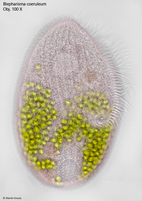
Fig. 2:Blepharisma coeruleum. L = 105 µm, ventral view with focus on the spherical macronucleus. Obj. 100 X.
Fig. 3:Blepharisma coeruleum. L = 105 µm, ventral view with focus on the rows of pink granula. Obj. 100 X.
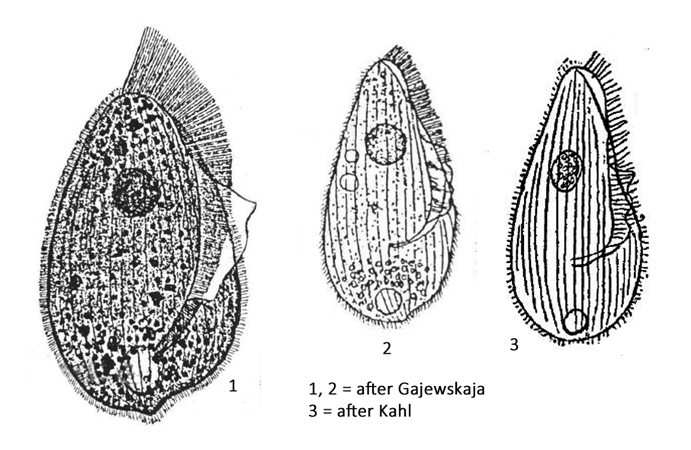
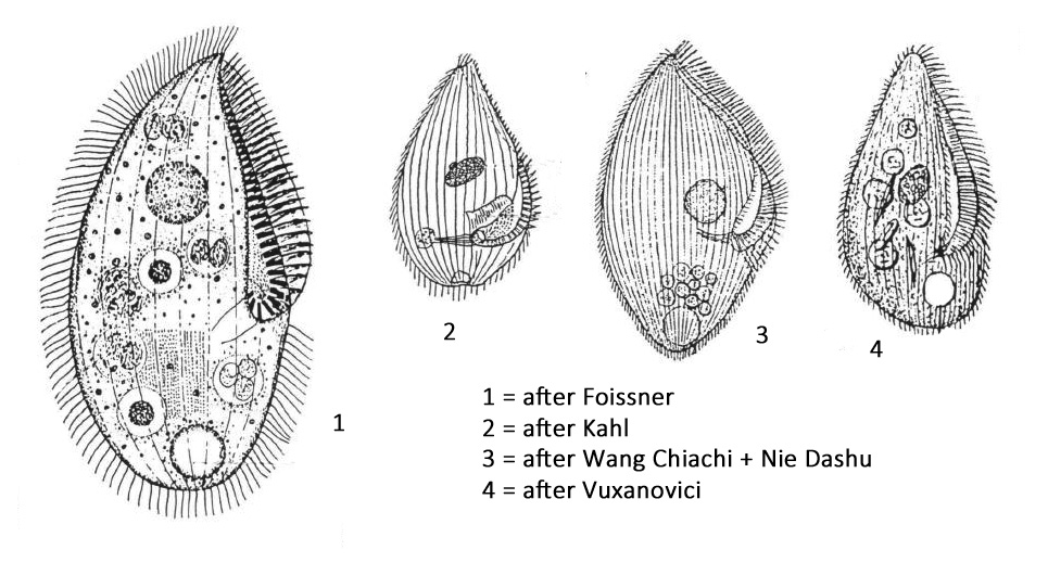
The anteriorly pointed shape and the pink coloration are very typical of Blepharisma. The length of the specimens observed varied between 100–120 µm, which is quite small for Blepharisma coeruleum. In fact, the specimens shown in Fig. 1 are very similar to Blepharisma lateritium, which is supposed to grow to a similar size:
Blepharisma lateritium
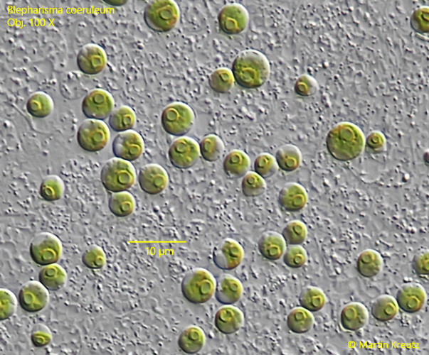
However, all of the specimens I have observed (about 40) contained symbiotic algae that filled the posterior half of the body. This is not described for Blepharisma lateritium. According to the original description of Blepharisma coeruleum by Gajevskaja this species should have a distinct bluish or blue-green coloration. This was not the case in my specimens, but the description of Gajevskaja was adopted by later authors (Kahl, Foissner) without having examined the species itself in vivo. Kahl pointed out the similarity between Blepharisma coeruleum and Blepharisma lateritium and also considers it a variant of Blepharisma lateritium. Kahl further writes (I assume according to Gajevskaja’s description): “apparently eats only one particular alga (Tetraspora)”. In fact, the algae in my specimens were all of the same species and look very similar to Tetraspora (s. Fig. 4). However, the algae are certainly not ingested, but symbiotic algae. I conclude this on the basis of the following observations:
– all observed specimens had about the same number of algae in the plasma, always in the posterior end – all algae are of the same species – the clearly visible food vacuoles of the specimens exclusively contained bacteria (s. Fig. 5) – the algae are not sequestered in separate vacuoles
Fig. 4:Blepharisma coeruleum. The symbiotic algae with a diameter of 4–6 µm are not of the Chlorella type but similar to Tetraspora. Obj. 100 X
The presence of symbiotic algae confirms the identification as Blepharisma coeruleum. However, there is some uncertainty regarding the state of the nuclear apparatus. Gajevskaja does not describe a number of micronuclei. For the similar species Blepharisma lateritium there are different data. Kahl indicates the presence of only one micronucleus in this species, while Foissner describes “many small” micronuclei. This may mean that the number of micronuclei in Blepharisma lateritium is variable. In my population specimens with 3–6 micronuclei were present:
Fig. 6:Blepharisma coeruleum. The macronucleus with 4 attached micronuclei. Mi = micronuclei, Ma, = macronucleus. Obj. 100 X.
A further uncertainity is the color of the granula. Gajevskaja described a blue-green coloration. Therefore I examined the ciliate again in brightfield, as Gajevskaja probably did:
Fig. 7:Blepharisma coeruleum. Color of the granula in bright field illumination is clearly pink. Obj. 100 X.
The image in Fig. 7 indicates clearly a pink color. However, if the aperture is closed a bit, they appear almost black. It is difficult to say under what conditions and with what kind of optical equipment Gajevskaja investigated the color of the granules at that time, but my specimens were definitely not blue or blue-green. It is known, however, that the dye in the granules (Blepharismin) can change color when exposed to light and oxygen. These facts demonstrate the difficulties in comparing present day observations with the descriptions and drawings of earlier authors. In my opinion, Blepharismacoeruleum with a pink coloration is present here, or Blepharisma lateritium with symbiotic algae. Because Blepharisma lateritium was found and described several times (in contrast to Blepharisma coeruleum) and symbiotic algae were never observed, I stick to the classification Blepharisma coeruleum.