body elongated oval, laterally flattened, posteriorly pointed
length 80–160 µm
adoral zone reaches mid-body
undulating membrane with half length of peristome
pellicle with bands of colorless cortical granules
macronucleus oval or oblong
2–7 micronuclei adjacent to macronucleus
contractile vacuole terminal
Blepharisma hyalinum
I found Blepharisma hyalinum in a moss sample that I had doused with tap water. After 2 weeks in a closed petri dish, a large population of Blepharisma haylinum had formed. However, I was unable to find any specimens in other moss samples prepared at the same time.
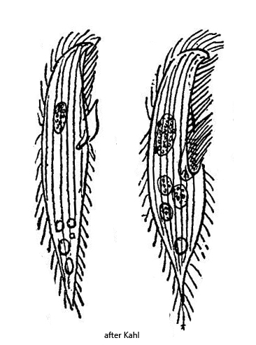
Kahl (1935) provides only a very brief description of Blepharisma hyalinum and two drawings (s. above). In 1983 the species was newly described by Larsen & Nilssen. The authors also carried out an examination of the colorless granules. The content of the granules shows an absorption maximum in the UV range. Components structurally related to blepharismin were not detected.
Blepharisma hyalinum is a rather small representative of the genus. Characteristic features are the beaked apical end and the pointed posterior end. The adoral zone extends right up to the middle of the body. The typical bands of granules are completely colorless in Blepharisma hyalinum. They also remain colorless when cultures are kept in the dark for years (Larsen & Nillson, 1983).
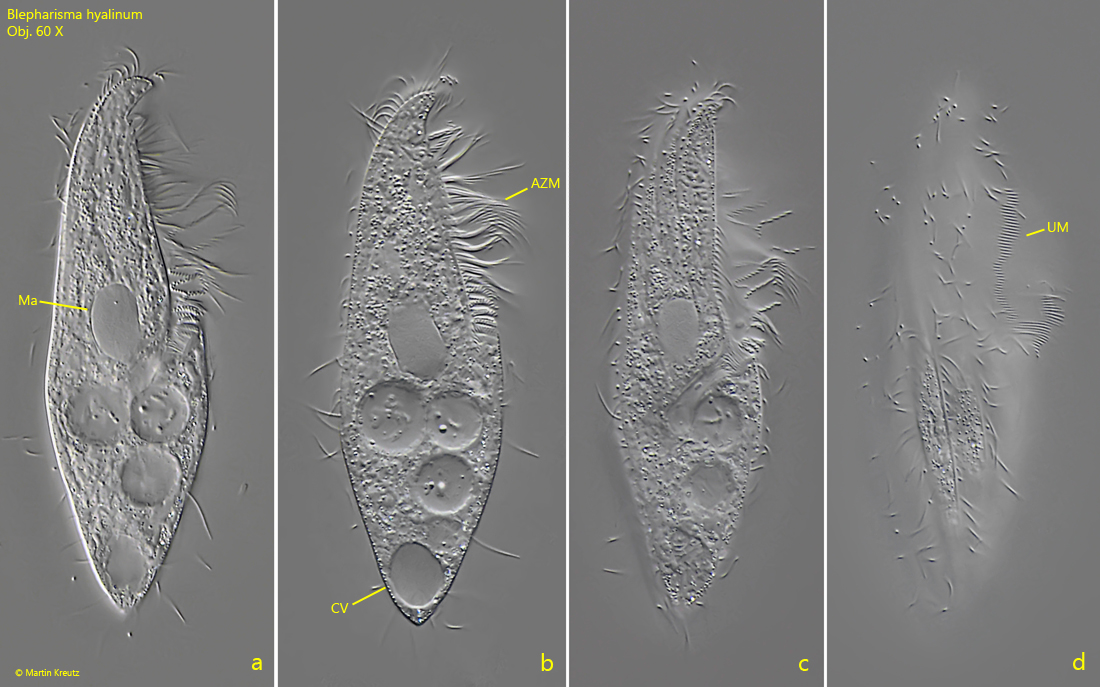
Fig. 1 a-d:Blepharisma hyalinum. L = 92 µm. Different focal planes of a freely swimming specimen from ventral. Note the pointed posterior end and the delicate undulating membrane (UM). AZM = adoral zone of membranelles, CV = contractile vacuole, Ma = macronucleus. Obj. 60 X.
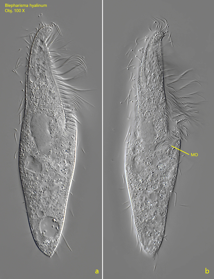
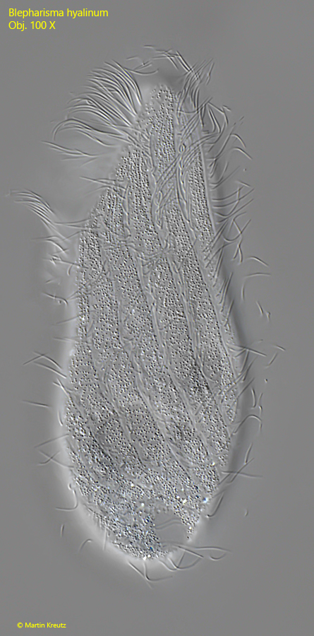
Fig. 2 a-d:Blepharisma hyalinum. L = 94 µm. A second freely swimming specimen from ventral. MO = mouth opening. Obj. 100 X.
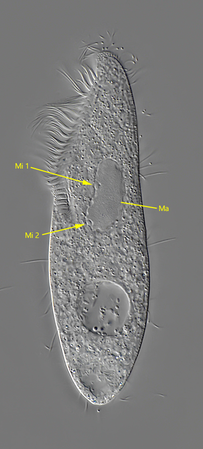
Fig. 3:Blepharisma hyalinum. A slightly squashed specimen from dorsal. Two of the four micronuclei (Mi 1, Mi 2) of this specimen are visible. Ma = macronucleus. Obj. 100 X.
Fig. 4:Blepharisma hyalinum. Focal plane on the bands of colorless cortical granules. Obj. 60 X.