body elongated oval, posterior end truncated transversely
length 80–180 µm
conspicuous paroral membrane parallel to the adoral zone on right side
adoral zone very broad, narrowing to the mouth opening in mid body
adoral zone running obliquely over ventral side, bent to left before mouth opening
contractile vacuole terminal
macronucleus oval or globular with adjacent spherical micronucleus
posterior end with long caudal cilia
Bothrostoma undulans
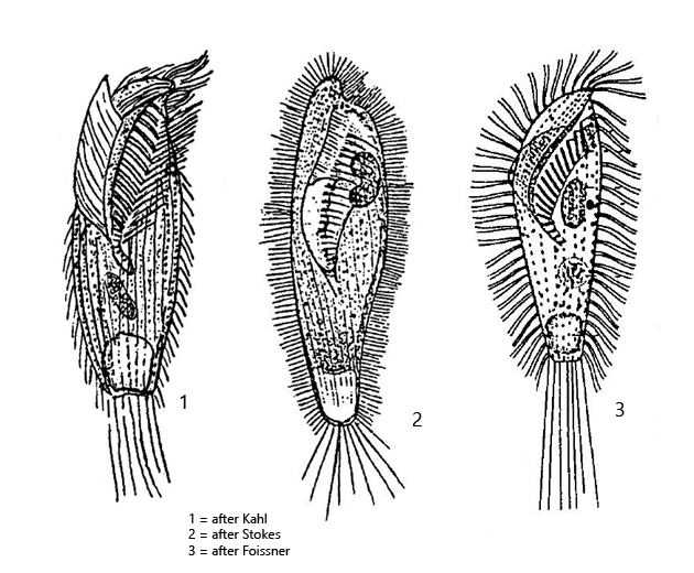
I find Bothrostoma undulans rarely but regularly in the Purren pond and the Simmelried, especially in decaying leaves. The ciliate is very conspicuous by its large, broad adoral zone (s. figs. 1 b and 2 a-c). This runs from left to right diagonally across the ventral side of the cell body and turns to the left just before the mouth opening (s. fig. 2 b). More difficult to recognize is the paroral membrane, which lies like a triangular sail over the adoral zone. The paroral membrane originates on the right side of the adoral zone and runs in parallel to it (s. fig. 2 c). Its complete shape is not easily seen, but the long cilia of the membrane lie like a curtain over the adoral zone in lateral view (s. figs. 1 a and 1 d). The macronucleus was also kidney-shaped in some specimens in my population (s. fig. 3) and enclosed partly the micronucleus. The length and numbers of caudal cilia also appear to be quite variable. I could also observe specimens with much longer caudal cilia than in the specimens shown below. The pellicle is clearly granulated by mucocysts, which are arranged in parallel stripes (s. fig. 4 b). In the cytoplasm I could detect symbiotic bacteria in all specimens, but I did not examine them more closely.
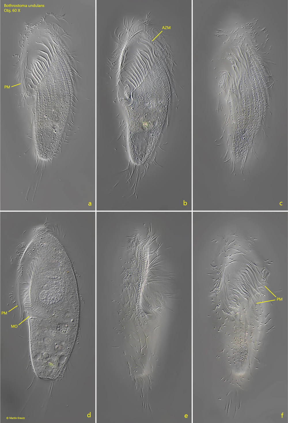
Fig. 1 a-f:Bothrostoma undulans. L = 96 µm. A freely swimming specimen from ventral (a, b, f), left (c, d) and right (e). Note the long cilia of the paroral membrane (PM) covering the adoral zone (AZM). MO = mouth opening. Obj. 60 X.
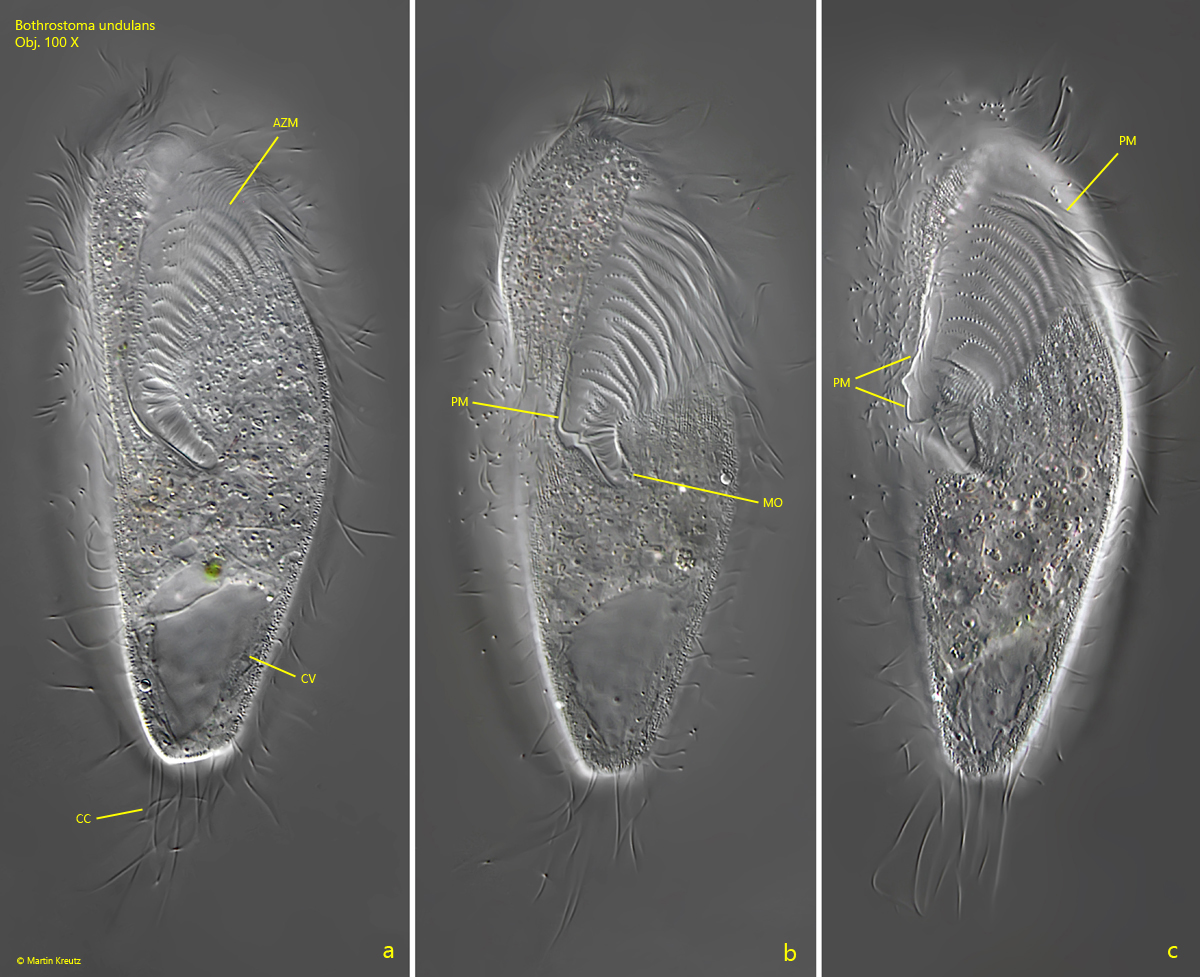
Fig. 2 a-c:Bothrostoma undulans. L = 114 µm. Three focal planes of a freely swimming specimen from ventral. AZM = adoral zone of membranelles, CC = caudal cilia, CV = contractile vacuole, MO = mouth opening, PM = paroral membrane. Obj. 100 X.
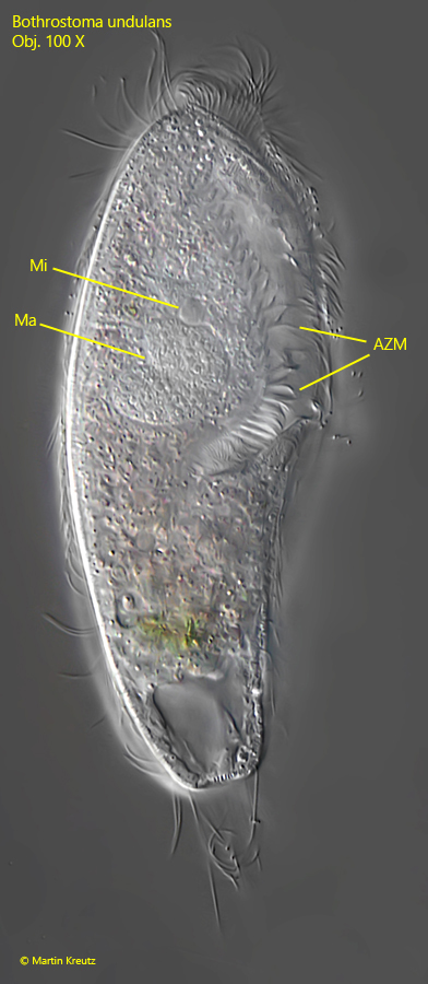
Fig. 3:Bothrostoma undulans. L = 114 µm. The same specimen shown in fig. 2 a-c from right. AZM = adoral zone of membranelles, Ma = macronucleus, Mi = micronucleus. Obj. 100 X.
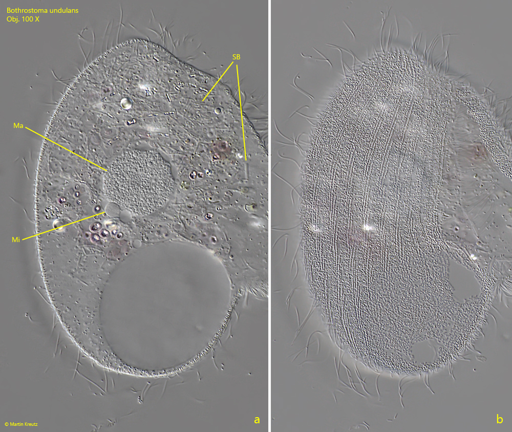
Fig. 4 a-b:Bothrostoma undulans. Two focal planes of the macronucleus (Ma) and micronucleus (Mi) and of the granulated pellicle (b) of a strongly squashed specimen. Inside the cytoplasm scattered symbiotic bacteria (SB) are visible. Obj. 100 X.