body plump pear-shaped, posteriorly caudate with blunt tip
apical dome hemispherical
length 60–80 µm
adoral zone runs equatorially and encircles body almost completely
mouth opening in posterior third
cytoplasm colorless, finely granulated
macronucleus elongated
contractile vacuole subterminal
tuft of caudalcilia, up to body length
Brachonella cydonia
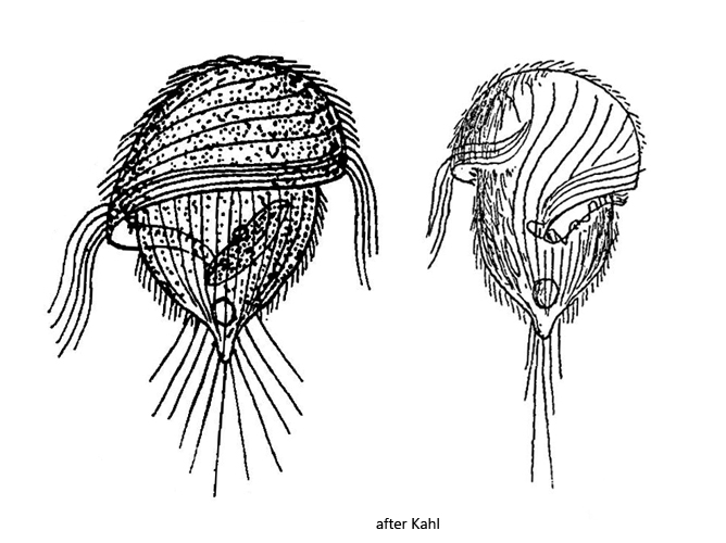
Brachonella cydonia first described as Metopus cydonia by Kahl in 1927. Later Jankowski (1964) transferred the species to the genus Brachonella. For the genus Brachonella, Jankowski defines the following characteristics:
– anterior end (dome) larger than the posterior end. – the adoral zone describes a helical line when it runs to the mouth opening
I find Brachonella cydonia rarely but regularly. So far all findings are from the Simmelried. The species is difficult to photograph, because of the high coverslip sensitivity. Already Kahl noted: “…kollabiert sofort beim Auflegen des Deckglases… (collapses immediately when the coverslip is put on)”. I can confirm this behavior, which is why I have not been able to examine any squashed specimen in detail. All specimens shown below were photographed at high layer thickness.
As the only deviation from Kahl’s description, I found the macronucleus to be often spherical, although Kahl himself was not sure if the elongated macronucleus is a constant feature. Kahl describes that the tuft of long caudal cilia can be actively spread. I have not been able to observe this with my specimens photographed swimming. Possibly this process can be seen only in resting specimens.
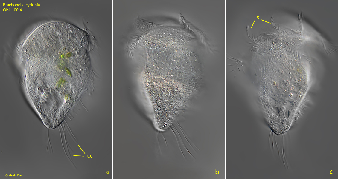
Fig. 1 a-c:Brachonella cydonia. L = 76. A freely swimming specimen. CC = caudal cilia, PC = perizonal cilia. Obj. 100 X.
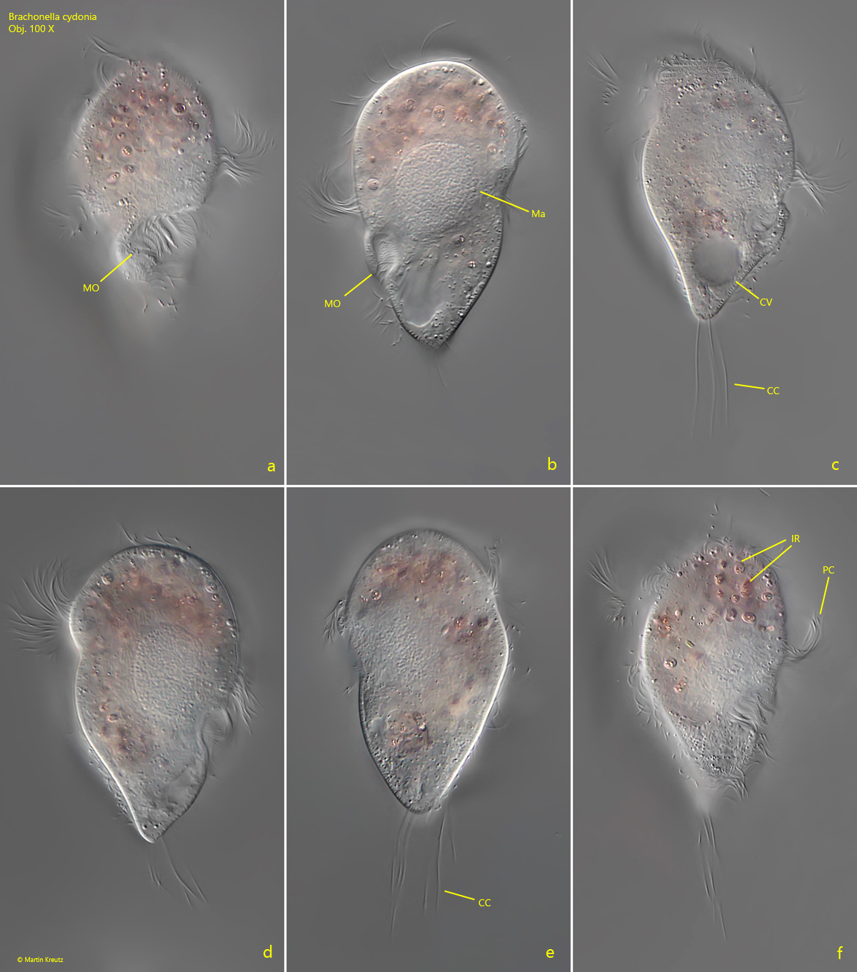
Fig. 2 a-f:Brachonella cydonia. L = 74. A second freely swimming specimen. CC = caudal cilia, CV = contractile vacuole, IR = ingested rhodobacteria, Ma = macronucleus, MO = mouth opening, PC = perizonal cilia. Obj. 100 X.
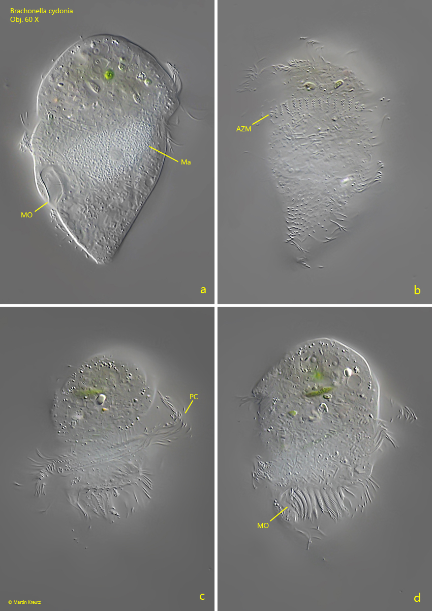
Fig. 3 a-d:Brachonella cydonia. L = 77. A damaged specimen that has already shed its caudal cilia due to coverslip pressure. AZM = adoral zone of membranelles, Ma = macronucleus, MO = mouth opening, PC = perizonal cilia. Obj. 100 X.