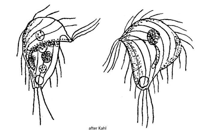
body broadly pyriform, apical dome overhangs left side in bead-shaped way
right margin of body merges into the apical dome without bulge
length about 50 µm
cilia long, flexible and widely spaced
adoral zone short with only 7–8 membranelles, limited to ventral side
perizonal stripe longer than adoral zone
globular macronucleus located in apical dome
one spherical micronucleus adjacent to the macronucleus
contractile vacuole large, terminal
long and delicate caudal cilia
Brachonella fastigata
Brachonella fastigata was first described in 1926 by Kahl as Metopus fastigatus. Later the species was transferred to the genus Brachonella by Jankowski (1964).
So far I could detect Brachonella fastigata exclusively in the Simmelried where the species is quiete common. Brachonella fastigata can be easily identified by the strongly overhanging bulge of the apical dome on the left side of the body. The right side of the body, on the other hand, is continuous with the apical dome without such a bulge. This gives the effect of a “peaked cap” in lateral view. Brachonella fastigata swims slowly with rowing movements of the long cilia. The very long, soft caudal cilia are also conspicuous. I had the impression that the posterior pole of the cell is free of cilia, which arise in a ring around it. The specimens in my population were 50 – 58 µm long, which is slightly larger than described by Kahl, who states “maximum 50 µm”. Otherwise all characteristics agree with the description of Kahl.
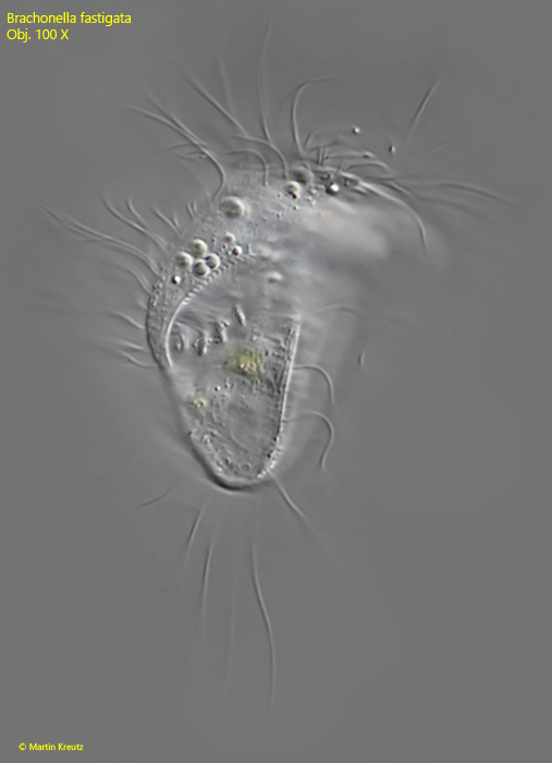
Fig. 1:Brachonella fastigata. L = 52 µm. A freely swimming specimen from ventral. Obj. 100 X.
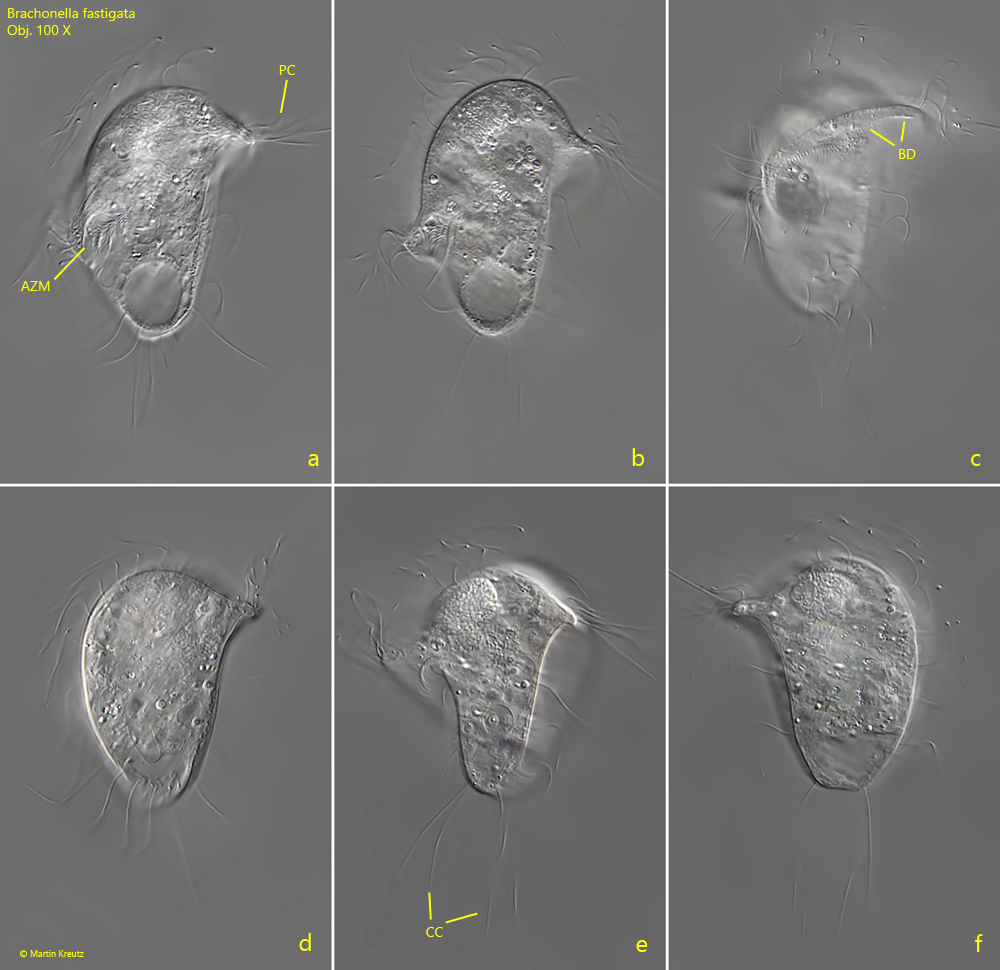
Fig. 2 a-f:Brachonella fastigata. L = 51 µm. A second freely swimming specimen from ventral (a, b, c, d), left (e) and from dorsal (f). Note the bulge of the apical dome (BD) hanging over the left side. AZM = adoral zone of membranelles, CC = caudal cilia, PC = perizonal cilia. Obj. 100 X.
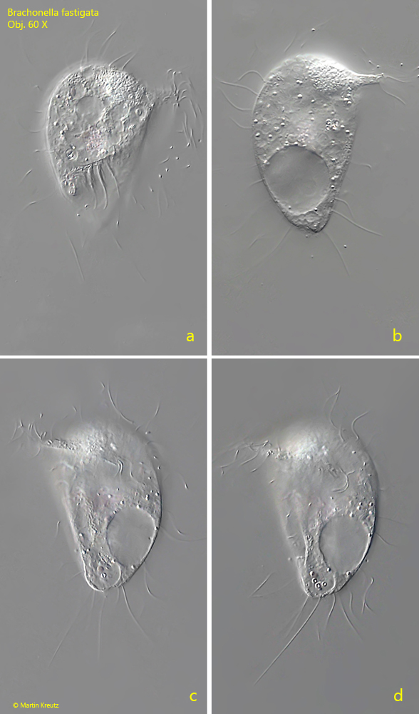
Fig. 3 a-d:Brachonella fastigata. L = 58 µm. A third freely swimming specimen from ventral (a, b) and dorsal (c, d). Obj. 60 X.
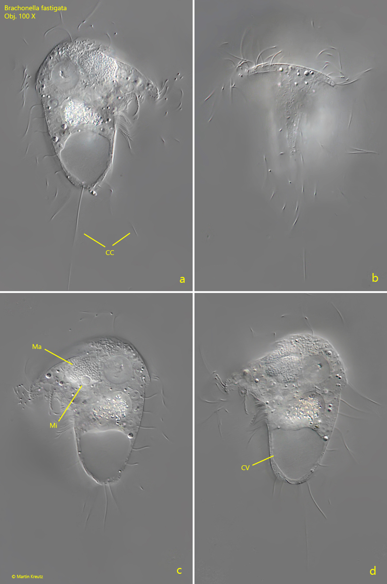
Fig. 4 a-d:Brachonella fastigata. L = 52 µm. A fourth freely swimming specimen from ventral (a), left (b) and dosal (c, d). CC =caudal cilia, CV = contractile vacuole, Ma = macronucleus, Mi = micronucleus. Obj. 100 X.
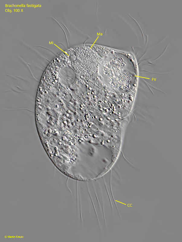
Fig. 5:Brachonella fastigata. A strongly squashed specimen. CC = caudal cilia, FV = food vacuole, Ma = macronucleus, Mi = micronucleus. Obj. 100 X.