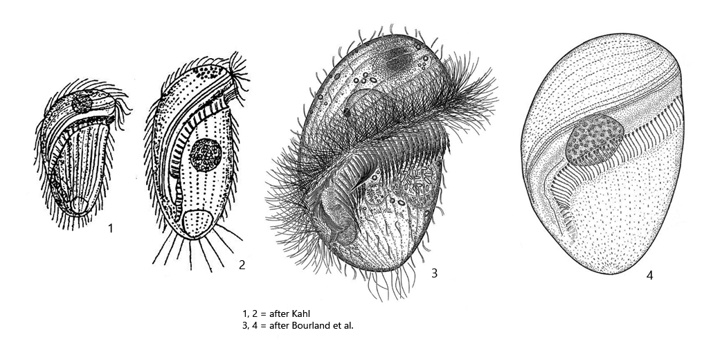
adoral zone running almost a full turn around longitudinal axis
on ventral side adoral zone runs diagonally bevor it bends to continue in parallel to longitudinal axis
mouth opening in posterior fourth of body
macronucleus globular
fringe of inconspicuous extrusomes beneath pellicle
contractile vacuole terminal
posterior end broadly rounded
slightly elongated caudal cilia
Brachonella pulchra
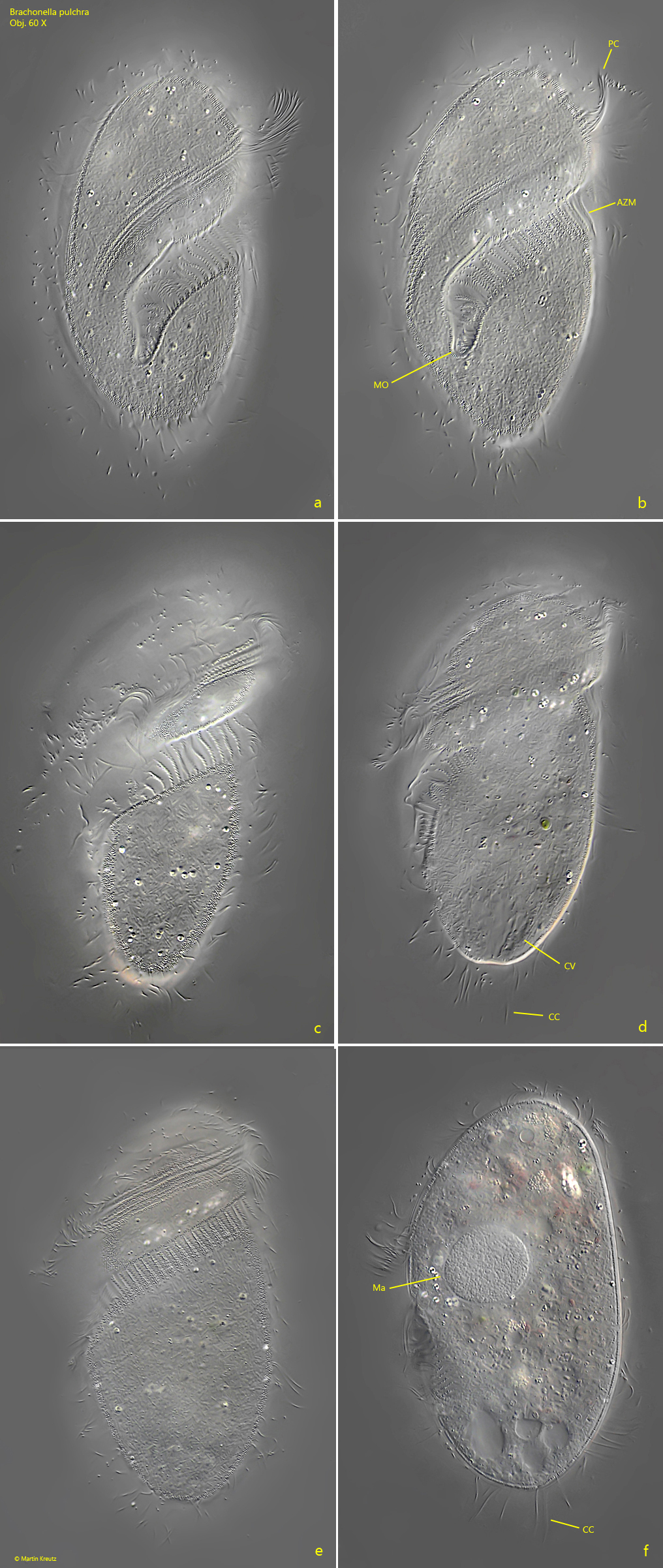
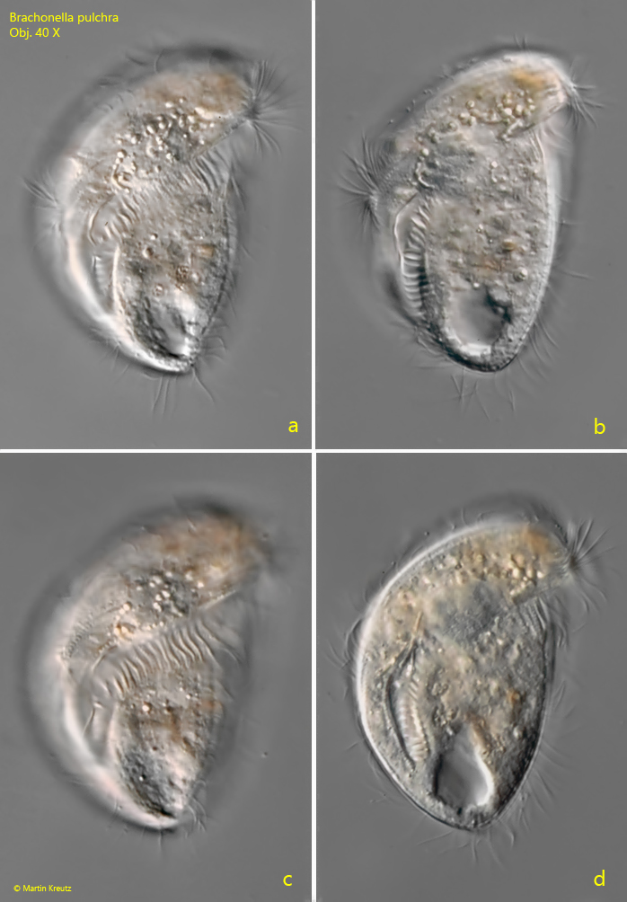
I have found Brachonella pulchraso far exclusively in the uppermost mud layer in Simmelried. The species is not very common. In the last 20 years I could observe only a few specimens. I recognize Brachonella pulchra by the very posteriorly located mouth opening (s. fig. 1 b) and the course of the adoral zone, which runs almost around the entire body and suddenly turns posteriorly on the ventral side, almost in parallel to the longitudinal axis. At the broadly rounded posterior end a few, slightly elongated caudal cilia arise, which can be easily overlooked. As already described by Kahl, the species is quite variable. So I could observe a smaller form in December 1998, which was only 84 µm long (s. fig. 2 a-d).
Fig. 1 a-f: Brachonella pulchra. L = 108 µm. A freely swimming specimen from ventral (a, b), from right (c, d) and from dorsal (e, f). AZM = adoral zone of membranelles, CC = caudal cilia, CV = contractile vacuole, Ma = macronucleus, MO = mouth opening, PC = perizonal cilia. Obj. 60 X
Fig. 2 a-d: Brachonella pulchra. L = 84 µm. A second, freely swimming specimen from ventral. Obj. 40 X