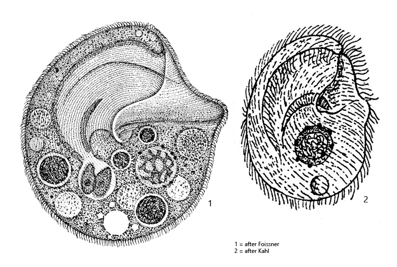
body broadly oval or almost circular, shape variable
deeply indented at the mouth opening
vestibular bottom lip-shaped
length 180–250 µm, sometimes up to 400 µm
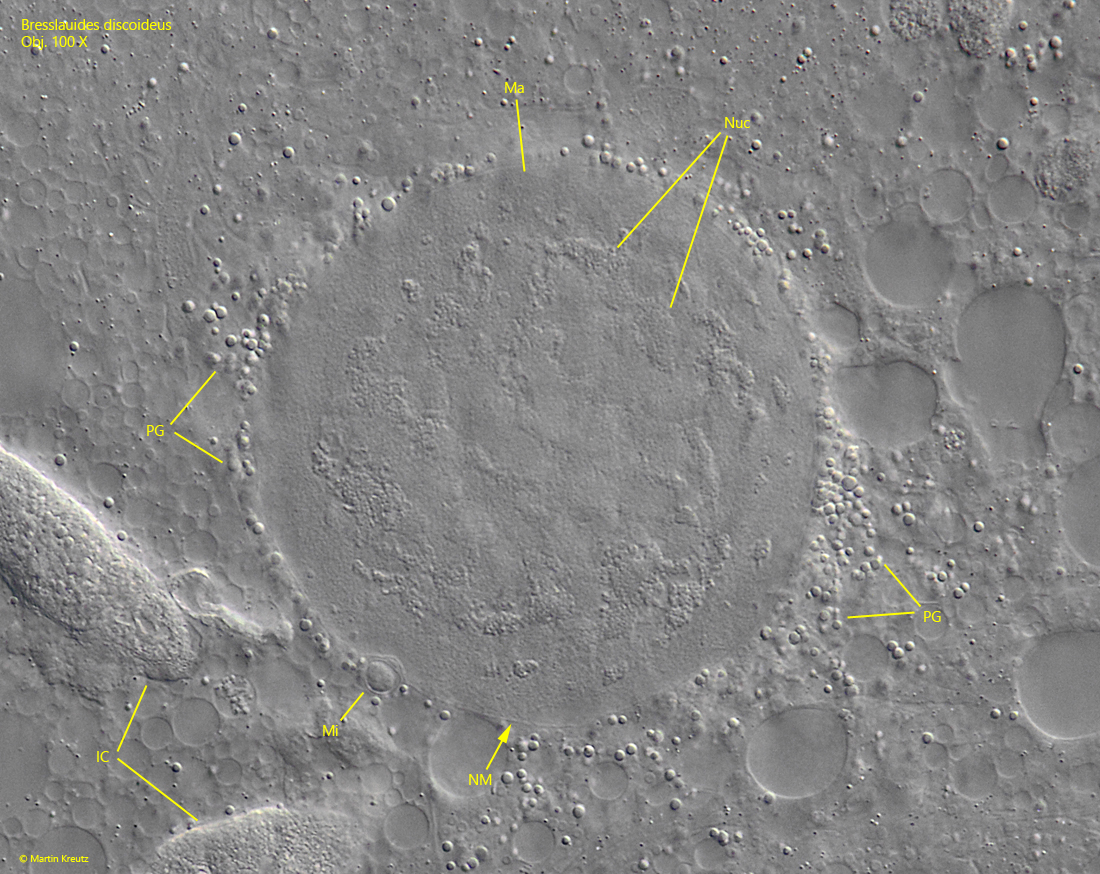
macronucleus globular to slighty ellipsoid, with reticulate nucleolus, nuclear membrane distinct
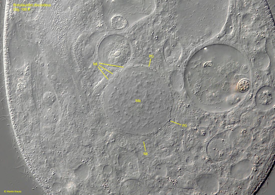
macronucleus surronded by a layer of perinuclear globules
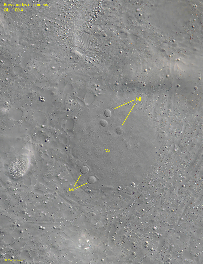
3–20 micronuclei adjacent to macronucleus, about 3–5 µm, surrounded by membrane
mouth opening with a right and left field of polykineties, right polykinetid thin and inconspicuous
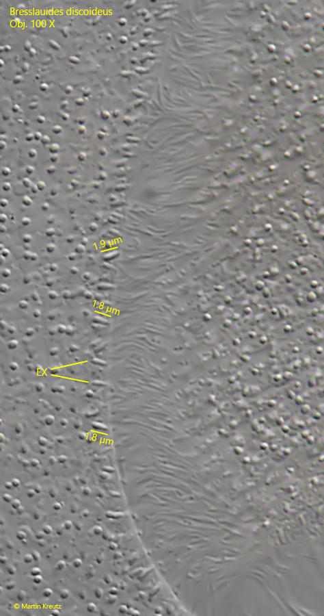
fusiform extrusomes 1.5–2.5 µm long
ciliary rows consiting of paired cilia
contractile vacuole subterminal
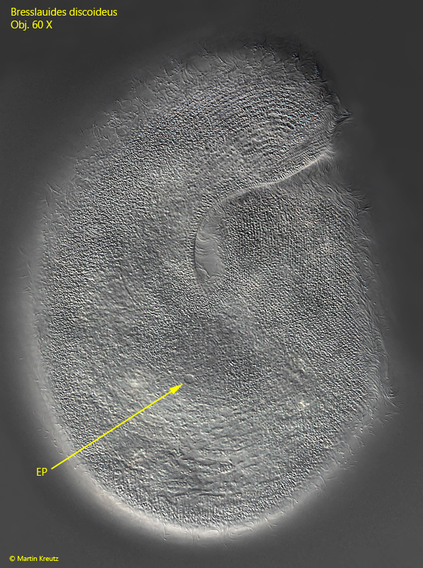
excretion porus in posterior third on right side
no caudal cilia
Bresslauides discoideus
I found my population of Bresslauides discoideus in moss samples, mainly from trees and walls. If the moos is poured over with rainwater in Petri dishes, Bresslauides discoideus can be found in some of the samples after a few days. Bresslauides discoideus has so far been found in mosses, soil samples and humus. Kahl found Bresslauides discoideus exclusively in moss samples.
Large populations of different ciliates develop in the moss samples after a few days. However, Bresslauides discoideus is immediately recognizable by its size and almost circular shape. In my population, the specimens were between 220–250 µm long. In addition, Bresslauides discoideus has a greatly enlarged vestibulum, with the lower half protruding like a lip. This gives the ciliate a typical shape in lateral view.
Bresslauides discoideus is feeding on other ciliates, but also rotifers and amoebae. To do this, it lies on its left side and swirl in the prey organisms. Due to this behavior, all specimens immediately turn to their left side when the samples is placed on the slide. This is why I have only managed to take images of the right side so far.
A characteristic feature of Bresslauides discoideus is a layer of perinuclear globules around the macronucleus. This layer can be differently pronounced. In some specimens the globules are arranged very dense and multilayered, while in other specimens the globules are only present in one layer. In addition, there are always several micronuclei adjacent to the macronucleus, which distinguishes the genus Bresslauides from the genera Colpoda and Bresslaua.
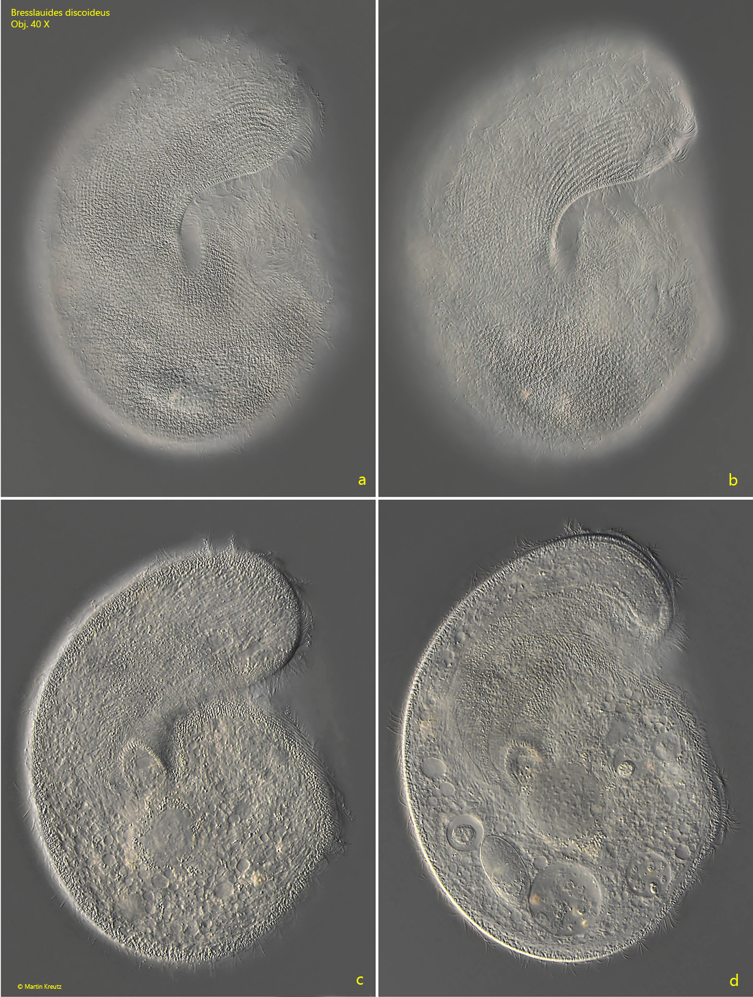
Fig. 1 a-d:Bresslauides discoideus. L = 232 µm. A freely swimming specimen from right. Obj. 40 X.
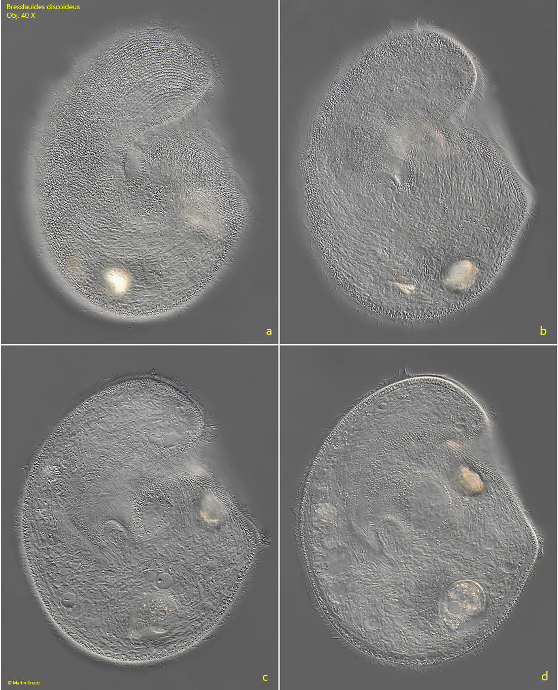
Fig. 2 a-d:Bresslauides discoideus. L = 225 µm. A second freely swimming specimen from right. Obj. 40 X.
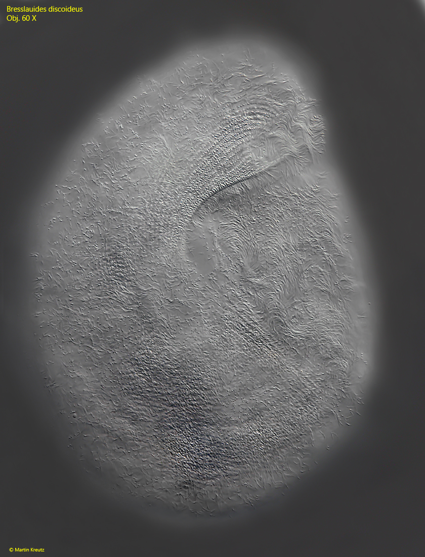
Fig. 3:Bresslauides discoideus. L = 245 µm. The somatic ciliation of the right side. Obj. 60 X.
Fig. 4:Bresslauides discoideus. The excretion porus (EP) on the right side is far away from the posterior end. Obj. 60 X.
Fig. 5:Bresslauides discoideus. The globular macronucleus has a distinct nuclear membrane (NM) and is surrounded by a layer of perinuclear globules (PG). The nucleolus (Nuc) is reticulate and appears cloudy. One micronucleus (Mi) in a separate membrane is visible. IC = ingested ciliates. Obj. 100 X.
Fig. 6:Bresslauides discoideus. The macronucleus of a second specimen with focal plane on the adjacent micronuclei (Mi). Each micronucleus is enveloped in a separate membrane. Obj. 100 X.
Fig. 7:Bresslauides discoideus. The macronucleus (Ma) with some adjacent micronuclei (Mi) and perinuclear globules (PG) in a third specimen. Obj. 60 X.
Fig. 8:Bresslauides discoideus. The extrusomes (EX) are fusiform and have in this specimen a length of 1.8–1.9 µm. Obj. 100 X.