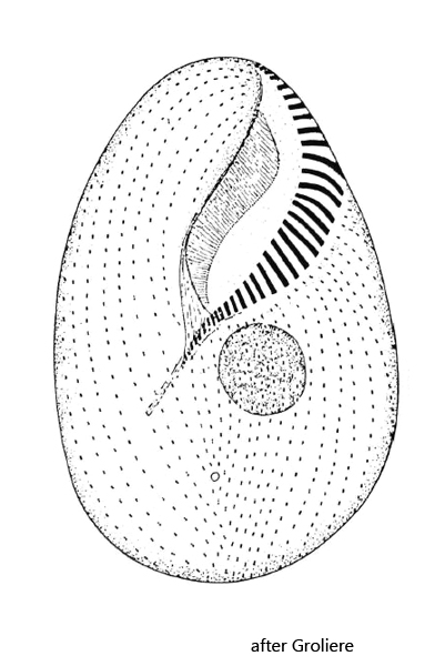
oral aperture in the upper left quadrant, oriented about 45° to longitudinal body axis
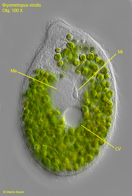
globular macronucleus with one adjacent micronucleus
contractile vacuole is located below the center of the cell
excretion pore of contractile vacuole in posteror third
70–80 somatic kineties
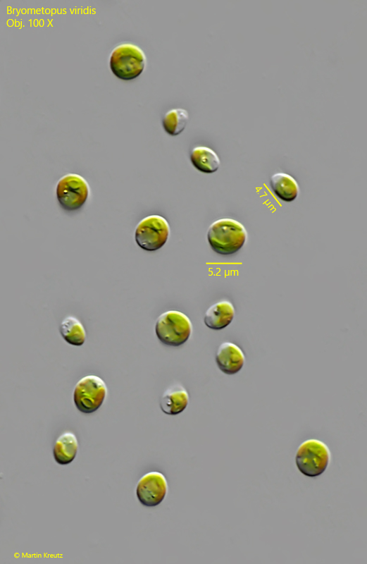
green due to symbiotic algae
Bryometopus viridis
I regularly find Bryometopus viridis in the Simmelried in squeezed Sphagnum moss and between floating leaves, which have not yet decomposed. The specimens swim after a short time already at the water surface, where they can be easily collected.
The locality is completely homogeneous with the locus classicus of this species, a bog near Besse-en-Chandesse in France, where Groliere also found the species in submerged Sphagnum moss in 1977. Foissner raised the green variant with symbiotic algae to species level in 1987. The variant without symbiotic algae is Bryometopus sphagni.
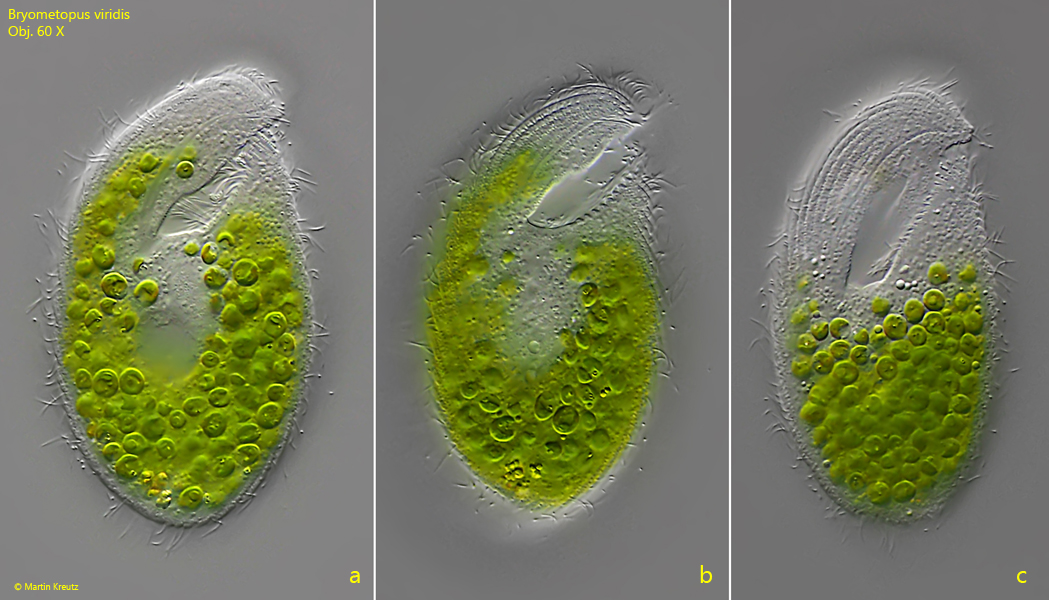
Fig. 1 a-c:Bryometopus viridis. L = 81 µm. Three focal planes of a freely swimming specimen. Obj. 60 X.
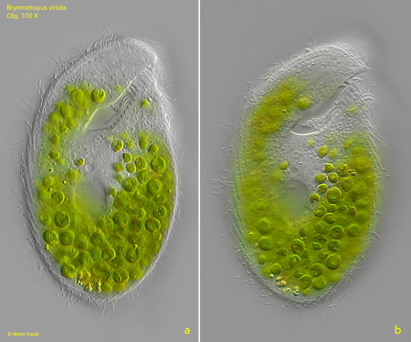
Fig. 2 a-b:Bryometopus viridis. L = 81 µm. Two focal planes of a slightly squashed specimen. Obj. 100 X.
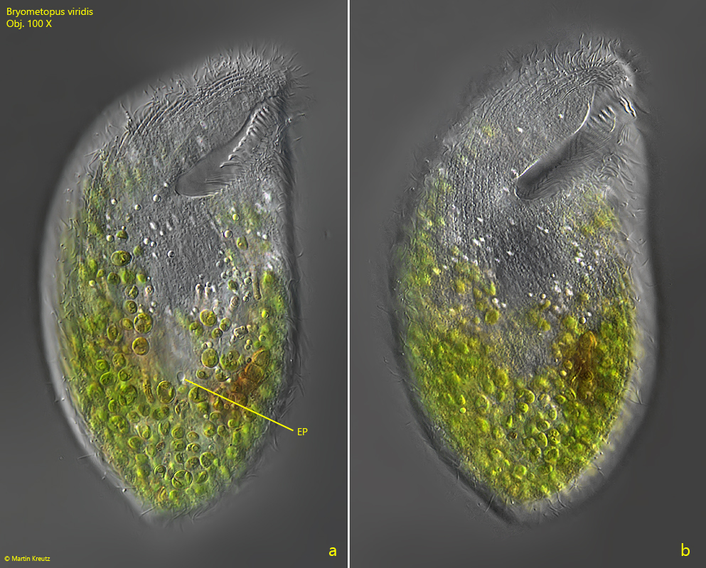
Fig. 3 a-b:Bryometopus viridis. L = 118 µm. Two focal planes of a second slightly squashed specimen. Note the excretion porus (EP) of the contractile vacuole in the posterior third of the cell (fig. 3a). Obj. 100 X.
Fig. 4:Bryometopus viridis. A squashed specimen for visualization of the globular macronucleus (Ma) with the adjacent micronucleus (Mi) and the contractile vacuole (CV) below the center of the cell. Obj. 100 X.
Fig. 5:Bryometopus viridis. The symbiotic algae in detail. Obj. 100 X.