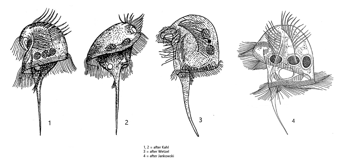
body medusoid with a prominent dome and a long caudal spine
length 80–200 µm
on dorsal side of the dome two rows of cirri (highly thigmotactic)
without somatic cilia except for a tuft of cilia at the base of the caudal spine
perizonal stripe consists of 5 ciliary rows
adoral zone runs in a furrow and encircles the body spirally
mouth opening in mid-body, cytopharynx directed anteriorly
2 or 3 macronuclei
one micronucleus
one contractile vacuole located at the base of caudal spine
Caenomorpha medusula
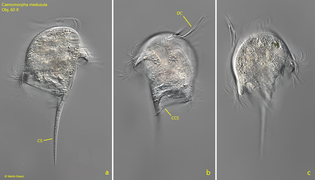
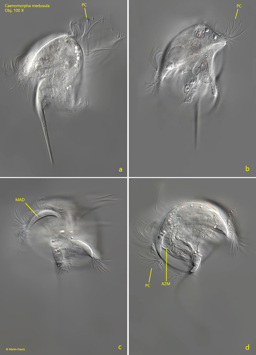
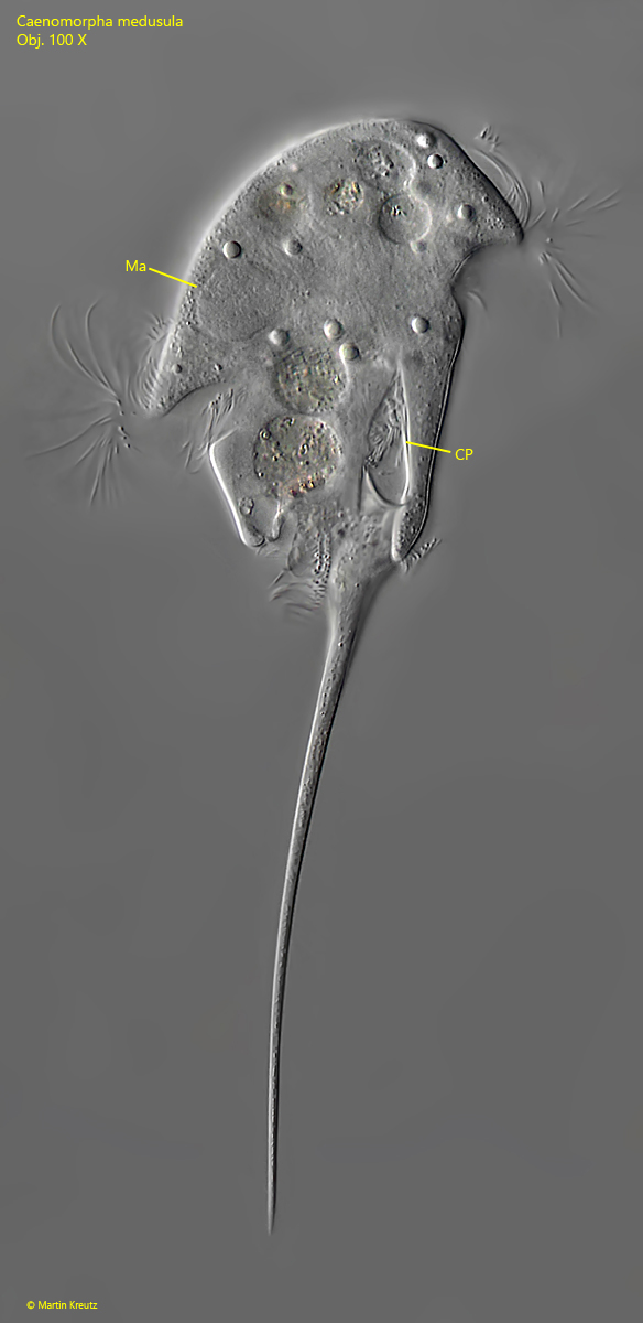
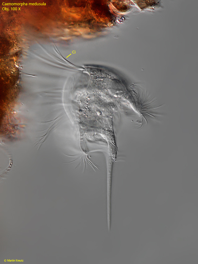
I find Caenomorpha medusula in all my sampling locations with a sapropelic mud layer of decaying leaves. Caenomorpha medusula is easily recognized by its typical shape, which resembles a jellyfish. The rigid caudal spine can reach different lengths according to my observations and the body length is also quite variable. This metopid ciliate is a fast swimmer, but it can hold on to the substrate thigmotactically with the dorsally located cirri (s. fig. 5). The cirri are surprisingly long. On attached specimens one can observe how the activity of the adoral zone and the perizonal cilia generates a water current to capture food particles (mainly bacteria). The mouth opening is located approximately in the middle of the body at the level of the contractile vacuole. However, the cytopharynx is directed towards the anterior end (s. fig 4). The adoral zone begins dorsally, to the left of the two rows of thigmotactic cirri and completely encircles the body to the mouth opening. Here, the adoral membranelles lie in a furrow covered by the overhanging margin of the apical dome. The perizonal stripe runs in parallel to the adoral zone up to the distal margin of the apical dome. At the base of the caudal spine a tuft of cilia is located (s. fig. 2b).
Fig. 1 a-c:Caenomorpha medusula. L = 104 µm. A freely swimming specimen from right (a), from left (b) and from dorsal (c). Obj. 60 X.
Fig. 2 a-c:Caenomorpha medusula. L = 127 µm. A second freely swimming specimen from right (a, b) and dorsal (b). Note the dosal rows of cirri (DC) and the tuft of cilia at the base of the caudal spine (CCS). CS = caudal spine. Obj. 60 X.
Fig. 3 a-d:Caenomorpha medusula. L = 105 µm. A third freely swimming specimen from left (a), right (b) and posterior (C, D). AZM = adoral zone of membranelles, MAD = margin of apical dome, PC = perizonal cilia. Obj. 100 X.
Fig. 4:Caenomorpha medusula. L = 170 µm. A fourth freely swimming specimen from right. CP = cytopharynx, Ma = macronucleus. Obj. 100 X.
Fig. 5:Caenomorpha medusula. L = 94 µm. This specimen holds onto a detritus flake with the thigmotactic dorsal cirri (Ci). Obj. 100 X.
Fig. 6 a-b:Caenomorpha medusula. L = 97 µm. A freely swimming specimen (a) and after attachement with the thigmotactic cirri (TC) to a detritus flake (b). Obj. 60 X.