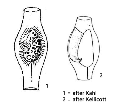
Calyptotricha pleuronemoides is described by Kahl as a rare species, of which he himself found only a few specimens. In my sampling sites Simmelried and Ulmisried I find it regularly in floating plants. The species is clearly identifiable because the ciliate builds a bulbous, transparent case, which is about 50 µm long. The ciliate itself is 22 – 35 long according to my measurements. However, this contradicts the data of Kahl, who gives a length of 85 µm for the lorica and “consistently 50 µm” for the ciliate. Whether or not the size of the specimens varies depending on the sampling site remains to be established. The loricae are usually attached to algal filaments. However, for observation at high magnifications, it is best to use the floating coverslip technique. Coverslips are carefully placed on the water surface of samples in which Calyptotricha pleuronemoides is present, where they float. After some days often some specimens of Calyptotricha pleuronemoides have settled on the coverslips. The loricae are then positioned immediately alongside the coverslip and can be easily observed without foreign bodies in the specimen.
The ciliate generates a continuous flow of water through the lorica by the constant activity of the sail-like undulating membrane. This draws in bacteria, flagellates, and small algae on which Calyptotricha pleuronemoides feeds. The movement of the undulating membrane also sets the ciliate into constant rotation.
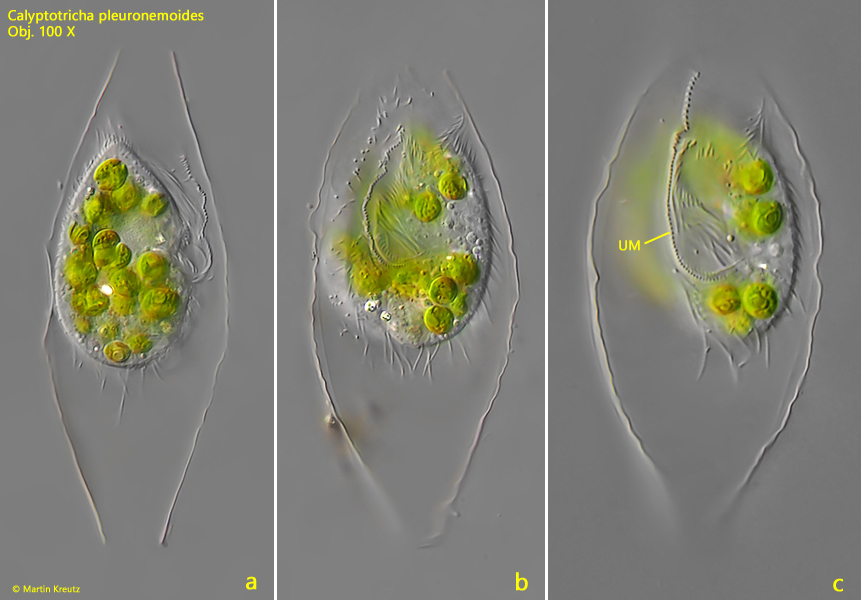
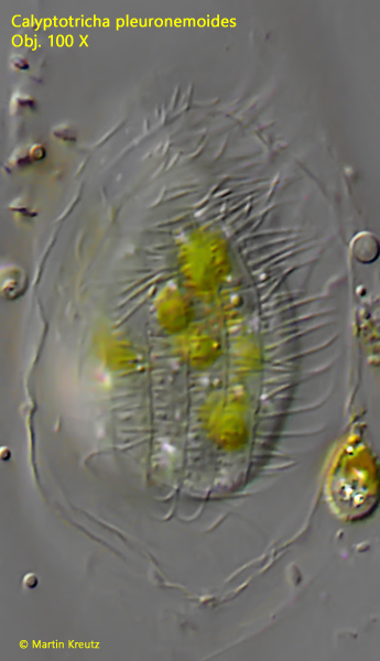
Fig. 1 a-c:Calyptotricha pleuronemoides. L = 33 µm. Three focal planes of an unsquashed specimen freely rotating in the lorica. UM = undulating membrane. Obj. 100 X.
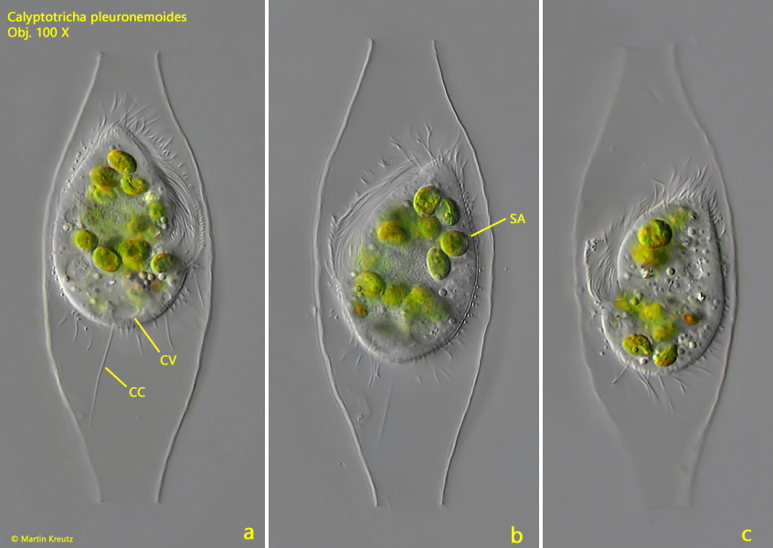
Fig. 2 a-c:Calyptotricha pleuronemoides. L = 35 µm. Three focal planes of a second specimen freely rotating in the lorica. CC = caudal cilium, CV = contractile vacuole, SA = symbiotic algae. Obj. 100 X.
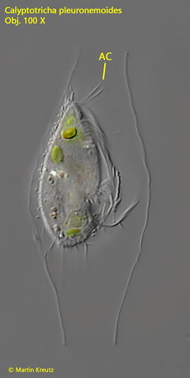
Calyptotricha pleuronemoides has two apical, elongated cilia, which probably have a tactile function. They are difficult to recognize in a rotating specimen in the lorica.
Fig. 3:Calyptotricha pleuronemoides. L = 30 µm. A specimen in lateral view with the two apical cilia (AC). Obj. 100 X.
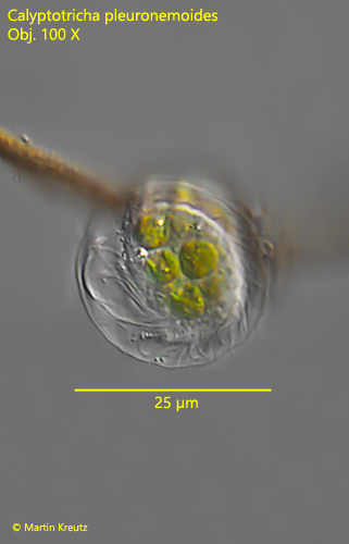
Fig. 4:Calyptotricha pleuronemoides. Apically view on a specimen rotating in the lorica. The circular lorica has a diameter of 25 µm. Obj. 100 X.
Fig. 5:Calyptotricha pleuronemoides. Focus on the dorsal ciliation. The kineties are arranged in distances of 3 µm. Obj. 100 X.
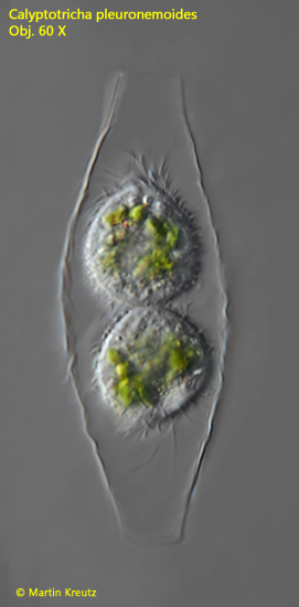
Fig. 6:Calyptotricha pleuronemoides. Two specimen in the lorica after cell division. Later one specimen will leave the lorica. Obj. 60 X.
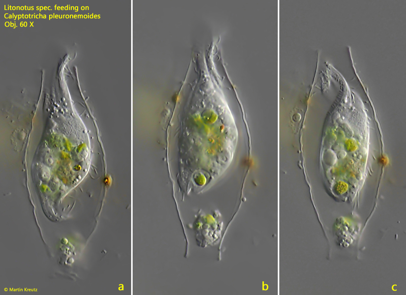
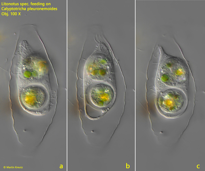
A predator of Calyptotricha pleuronemoides are the haptorian ciliates of the genus Litonotus. I could often find loricae invaded by small representatives of Litonotus (e.g. Litonotus alpestris or Litonotus lamella) feeding on Calyptotricha. Sometimes the specimens of Litonotus spec. were not able to leave the lorica due to the ingested prey, because they could not pass the lorica apertures. I assume that after digestion a cell division in the lorica took place.
Fig. 7 a-c: A specimen of Litonotus has entered the lorica and ingested Calyptotricha pleuronemoides. In the lower aperture of the lorica the excreted digest remains of Calyptotricha pleuronemoides can be seen. Obj. 60 X.
Fig. 8 a-c: This specimen of Litonotus spec. has ingested two specimens of Calyptotricha pleuronemoides and is unable to leave the lorica through the tight apertures. Obj. 100 X.