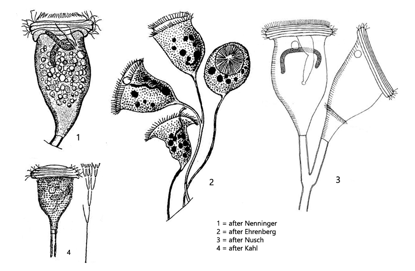
plasm colorless or yellow, filled with food vacuoles
peristome with 3–7 ciliary rows
one contracile vacuole
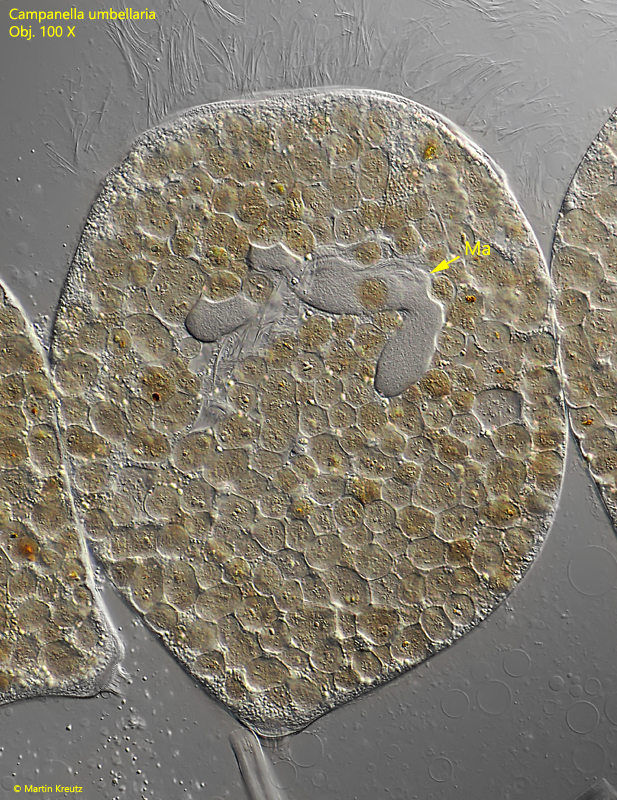
macronucleus horseshoe-shaped in anterior half of cell
pellicle covered with rows of rectangular or spherical tubercles
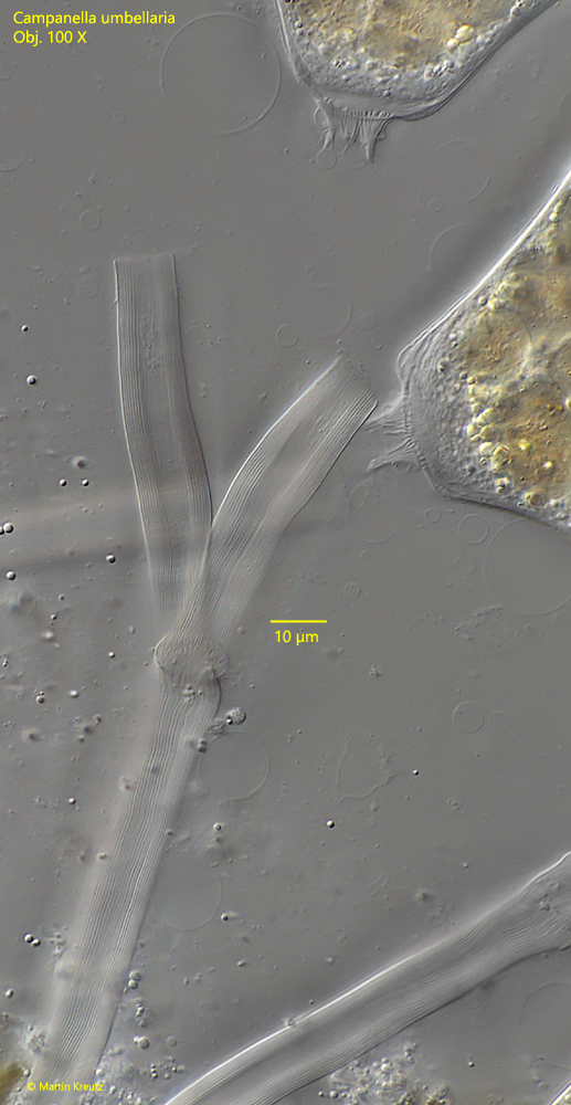
dichotomous, non-contractile stalk.
colonial
Campanella umbellaria
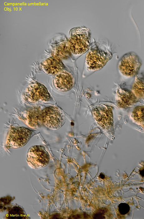
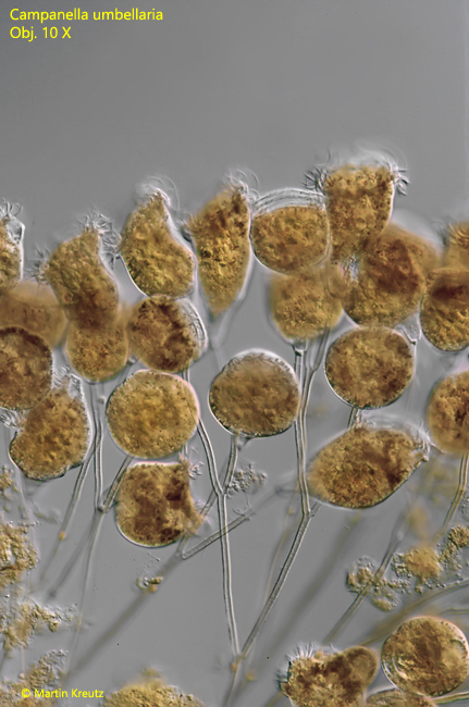
Campanella umbellaria is one of the most common peritrichous ciliates. Colonies are often found en masse as furry coverings on decaying leaves or plant stems. The species forms large colonies on branched stems. As an important distinguishing feature from Vorticella or Pseudovorticella, the stems are non-contractile. Only the individuals can contract. The individuals are very large, 200–300 µm, and usually completely filled with yellowish or brownish food vacuoles. As another distinctive feature Campanella umbellaria has a peristome with 3 to a maximum of 7 rows of cilia. The high number of cilia rows is already well visible in lateral view at low magnification.
Fig. 1, 2: Campanella umbellaria. L = 190–250 µm. Overview of two colonies at low magnification. Obj. 10 X.
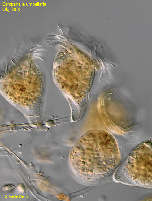
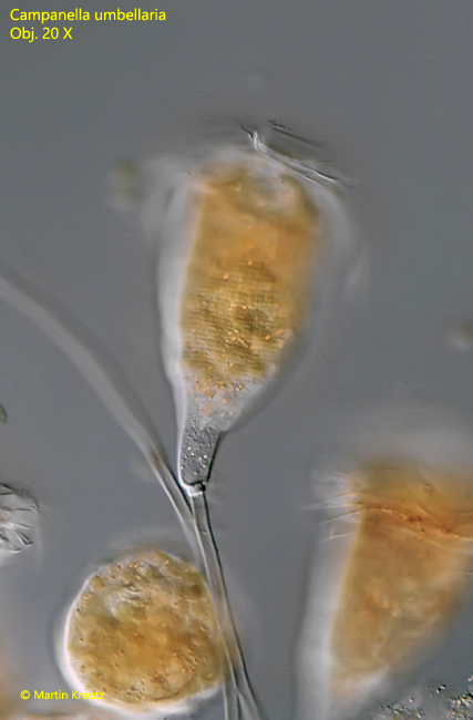
Fig. 3: Campanella umbellaria. L = 205 µm. An extended individuum in a colony. Obj. 20 X.
Fig. 4: Campanella umbellaria. L = 190–230 µm. An extended specimen with focal plane on the fine, transverse striation of the pellicle. Obj. 40 X.
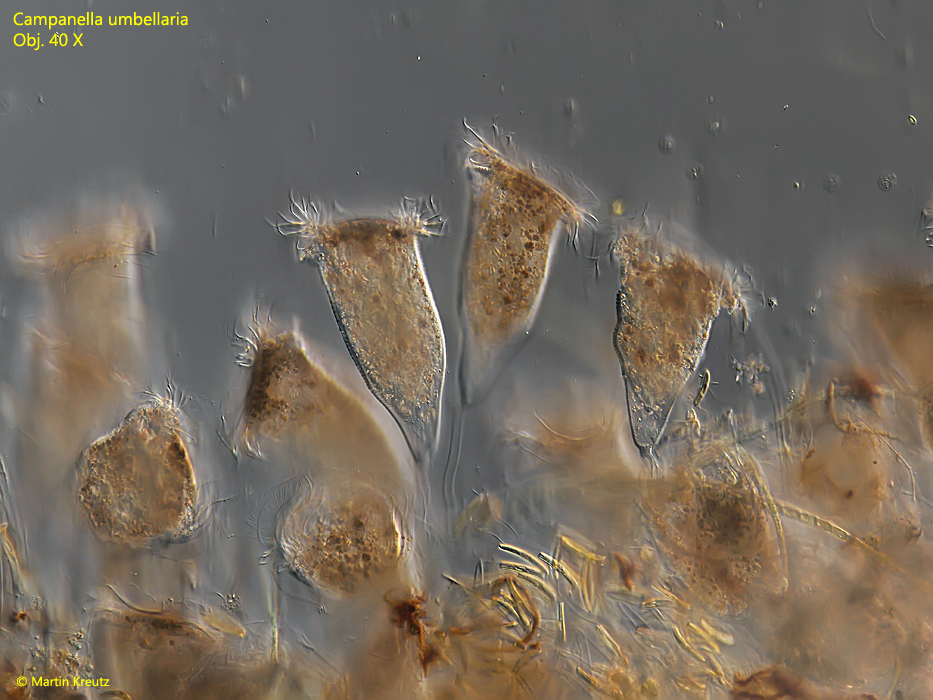
Fig. 5: Campanella umbellaria. L = 220–240 µm. Some extended individuals in a colony. Obj. 40 X.
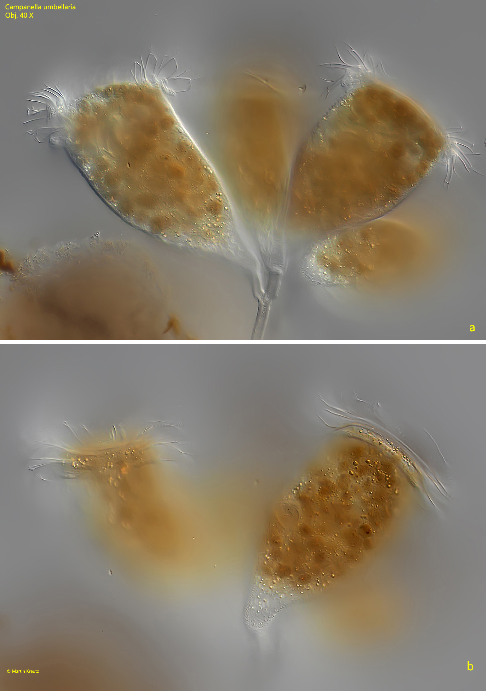
Fig. 6 a -b: Campanella umbellaria. L = 240 µm. Two focal planes of some extended individuals in a colony. Obj. 40 X.
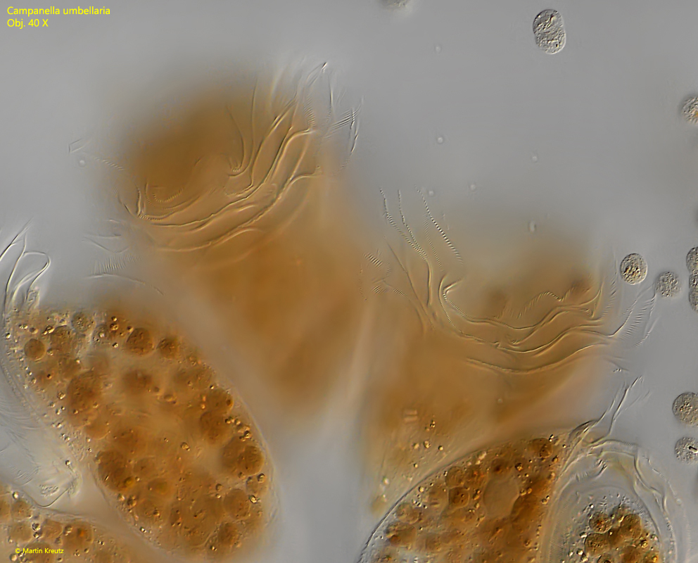
Fig. 7: Campanella umbellaria. Focus on the peristomial ciliary rows of some extended individuals. Obj. 40 X.
Fig. 8: Campanella umbellaria. The peristomial ciliary rows of a strongly squashed specimen. In this case the individuum has 5 of them. Obj. 100 X.
Fig. 9: Campanella umbellaria. Due to the large number of food vacuoles details in the cells are hard to recognize. In this strongly squashed specimen the horseshoe-shaped macronucleus is visible (Ma). Obj. 100 X.
Fig. 10: Campanella umbellaria. Detail of the non-contractile stalk with a diameter of 12 µm. Note the parallel striation and that the stalk is hollow. In the upper right edge of the image two detached individuals are visible. Obj. 100 X.