body elongated oval, posterior end broadly rounded
apical dome flat, not overhanging the left side
length 180–300 µm
adoral zone arises dorsally and runs obliquely to right side
perizonal stripe shorter than adoral zone
mouth opening at mid-body
about 90 narrowly spaced longitudinal kineties
macronucleus oval or dumbbell-shaped with adjacent micronucleus
cortex thick (about 3 µm)
some scattered caudal cilia (sometimes absent)
contractile vacuole terminal, large, with auxillary canals
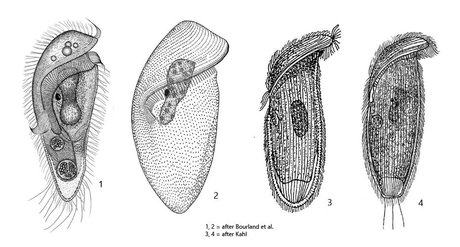
Castula fusca
Castula fusca was first described by Kahl in 1927 as Metopus fuscus. In 2014 Bourland et al. redescribed the species and in 2020 Bourland et al. transferred Metopus fuscus to the newly established genus Castula based on morphological characters and the results of a 18S rRNA gene analysis.
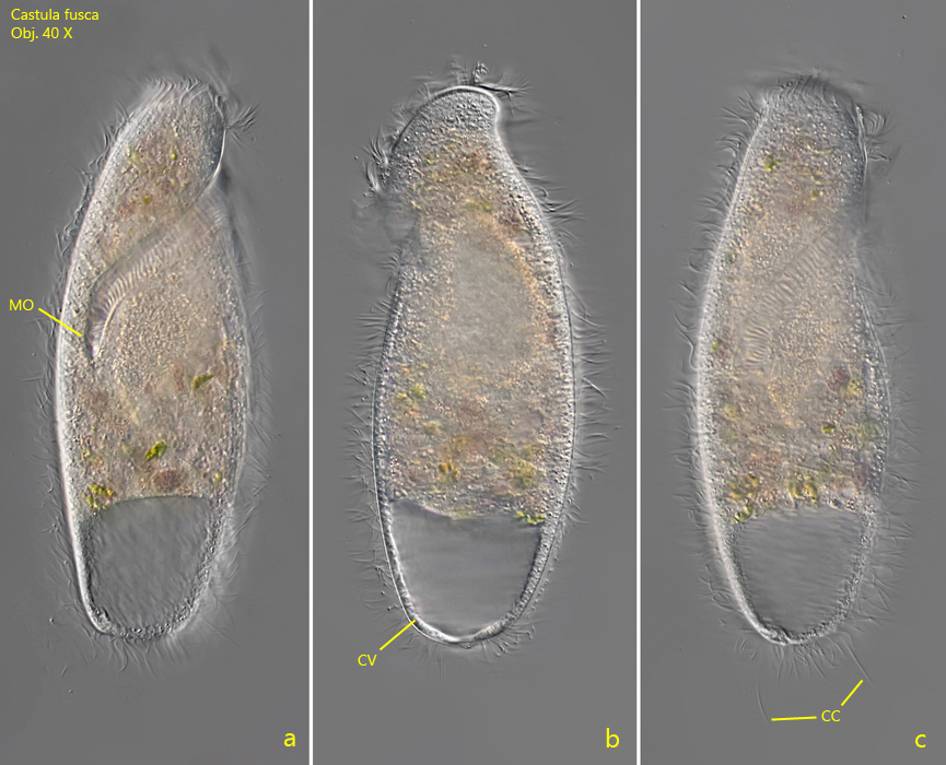
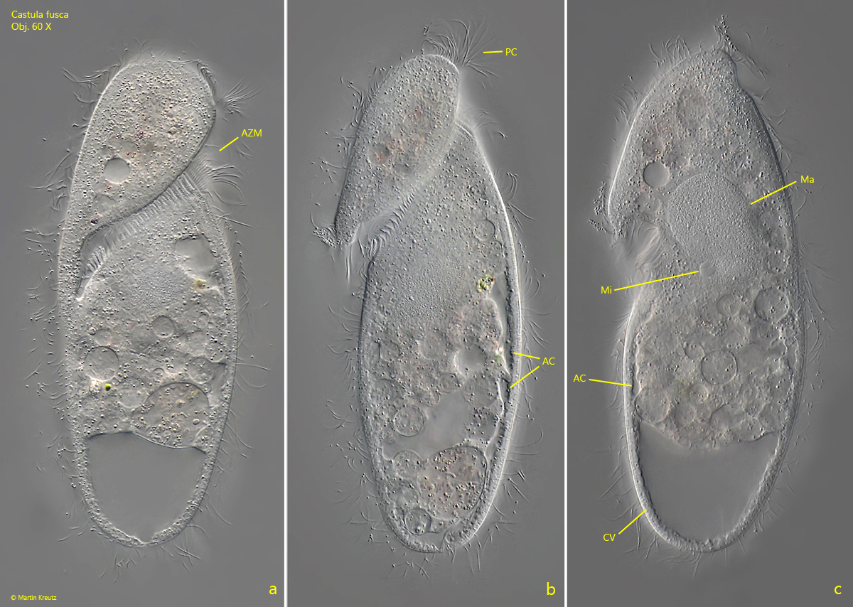
Castula fusca is the largest representative among the metopid ciliates and therefore easy to recognize even at low magnifications. I regularly find specimens in the Simmelried and the Purren pond. Mostly the specimens are brown or orange-brown and filled with food vacuoles. Only rarely transparent specimens can be found. At the posterior end Castula fusca has loosely arranged caudal cilia (s. fig. 1 c), but they can be easily overlooked. According to Kahl they can sometimes be absent. In the posterior half of the body there are auxiliary canals under the pellicle, which extend from the contractile vacuole to the level of the mouth opening in mid-body. These are also explicitly mentioned by Kahl. However, these canals were interpreted differently by Bourland et al. in the context of their redescription of Castula fusca in 2014. According to these authors, they are supposed to be flattened extensions of the contractile vacuole, which are formed when it is depressed from above by large food vacuoles. Bourland et al. assume this to be more an effect of the deformation of the contractile vacuole rather than real drainage canals. In my opinion, the length of these canals, which extend to the middle of the body, speaks against this interpretation. However, I have not checked whether these canals are a constant feature and whether they disappear in starving specimens with few food vacuoles.
Fig. 1 a-c:Castula fusca. L = 220 µm. A freely swimming specimen from right (a, c) and from left (b). Note the scattered caudal cila (CC) at the broadly rounded posterior end. CV = contractile vacuole, MO = mouth opening. Obj. 40 X.
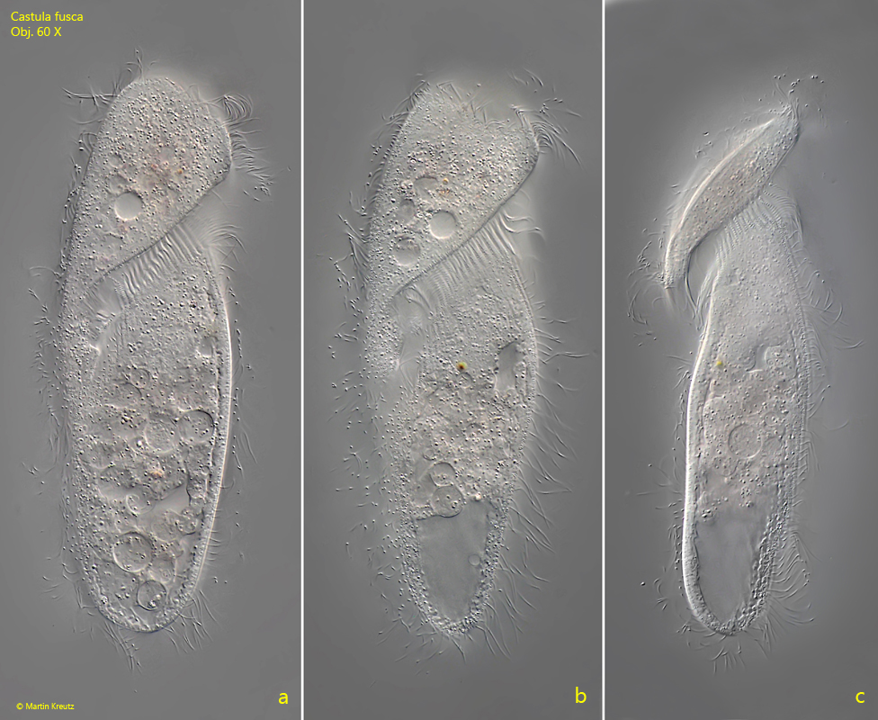
Fig. 2 a-c:Castula fusca. L = 184 µm. A second freely swimming specimen from right (a, b) and from left (c). Obj. 60 X.
Fig. 3 a-c:Castulafusca. L = 184 µm. The same specimen shown in fig. 2 a-c slightly squashed from right (a) and from left (b, c). Note the auxiliary canals (AC) of the contractile vacuole (CV). AZM = adoral zone of membranelles, Ma = macronucleus, Mi = micronucleus, PC = perizonal cilia. Obj. 60 X.