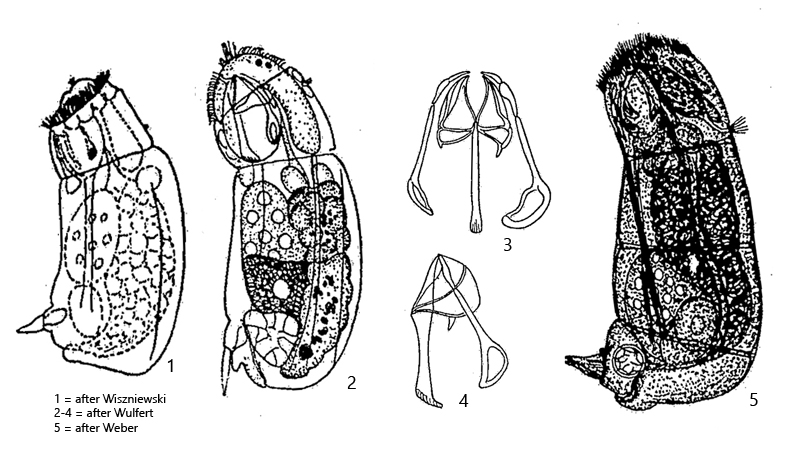
body stout, dorsally curved, ventrally almost straight
large head, distinct fold in neck
oblique corona without lips
cuticle flexibel
length 90–160 µm (with toes)
toes short, conical shaped, shifted to ventral side
two frontal eyespots close together (visible in dorsal view)
cerebral ganglion large and saccate
retrocerebral organ absent
trophi large, distal ends of manubria loop shaped
Cephalodella catellina
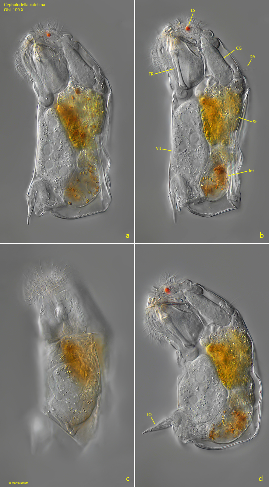
Up to now I have found Cephalodella catellina exclusively in the Bussenried and only a few specimens there. The specimen shown below was found in sample taken in November 2018. It was swimming near the surface of the sample.
The shape of Cephalodella catellina is very characteristic due to the short toes shifted to the ventral side. The posterior end is almost rectangular in shape. Due to the flexible cuticle, the shape is not constant and longitudinal and transverse folds are formed. Apically there are two eyespots that are close together. In lateral view, however, they appear as one eyespot.
Fig. 1 a-d:Cephalodella catellina. L = 110 µm (with toes). Different focal planes of a slightly squashed specimen. Note the ventrally shifted toes (TO). CG = cerebral ganglion, DA = dorsal antenna, ES = eyespot, Int = intestine, St = stomach, TR = trophi, Vit = vitellarium Obj. 100 X.