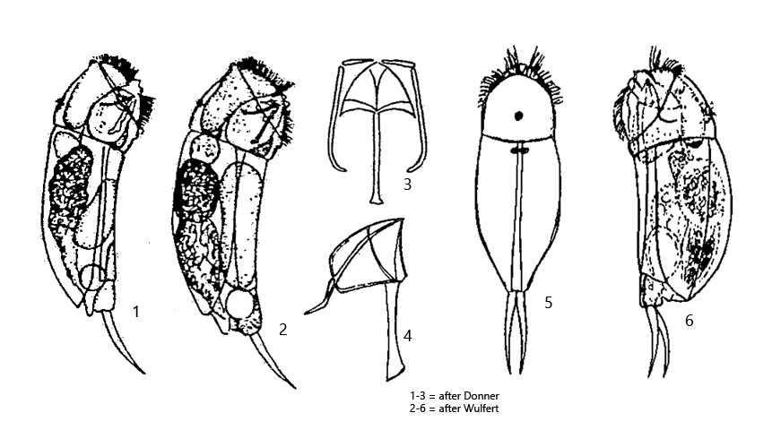
body stout, dorsally curved, ventrally almost straight
length 90–132 µm (with toes)
head narrower than body
end of fulcrum is bent upwards
lateral cleft widened posteriorly
corona oblique without lips
ganglion large and saccate with two eyespots at posterior end (visible in dorsal view)
retrocerebral organ absent
toes thin (20–32 µm), slightly ventrally curved with pointed ends
foot short and conical shaped
caudal antenna present
Cephalodella exigua
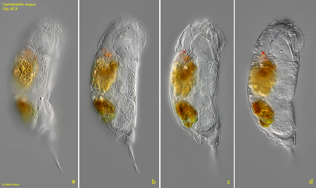
So far I have found Cephalodella exigua in the Bussenried and in the Simmelried in samples of decomposing plant material. I recognize Cephalodella exigua mainly by the thin toes, which are slightly curved ventrally and pointed at the end. The two cervical eyespots (appearing as one eyespot in lateral view) are located on the cerebral ganglion, which has an almost rectangular shape (s. fig. 2 a-b). The corona is clearly oblique and the stomach is golden brown or orange-brown in color. The stomach of the similar species Cephalodella auriculata is colored in the same way, but can be distinguished from Cephalodella exigua by its stout body shape and the broader head. In addition, the toes of Cephalodella auriculata are more curved.
Fig. 1 a-d:Cephalodella exigua. L = 130 µm (with toes). A freely swimming specimen from right. Obj. 60 X.
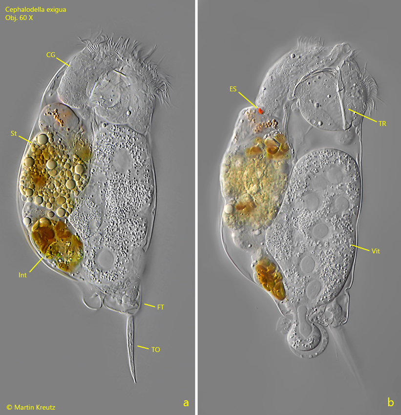
Fig. 2 a-b:Cephalodella exigua. L = 130 µm (with toes). The squashed specimen as shown in fig. 1 a-d. CG = cerebral ganglion, ES = eyespot, FT = foot, Int =intestine, St = stomach, TO = toes, TR = trophi, Vit = vitellarium. Obj. 60 X.
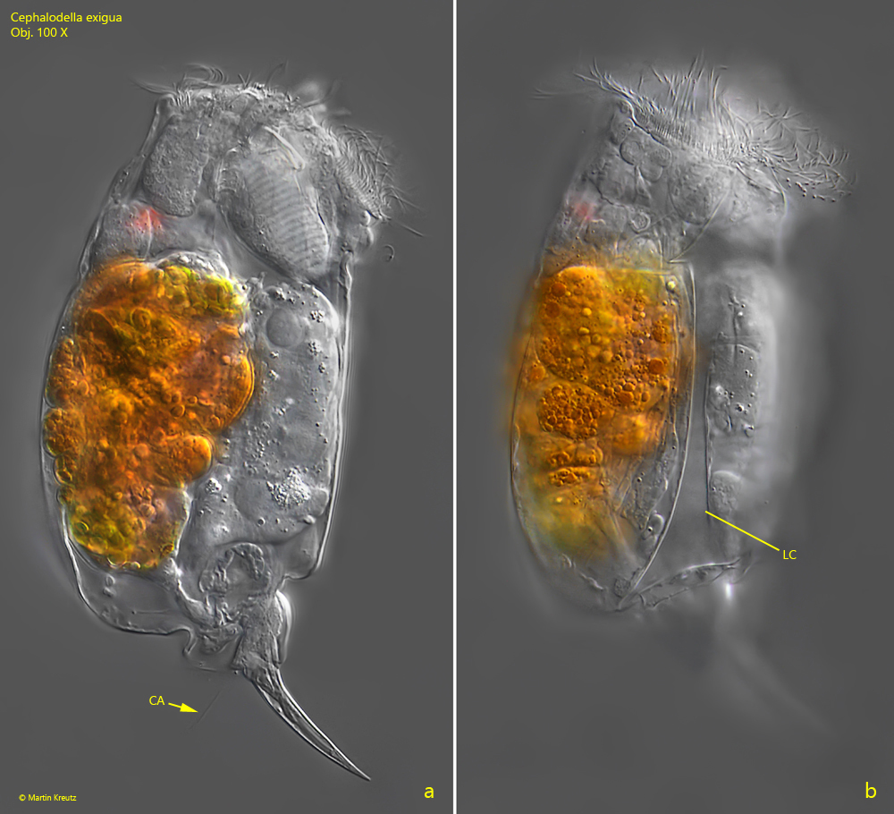
Fig. 3 a-b:Cephalodella exigua. L = 132 µm (with toes). A second, slightly squashed specimen from right. Note the caudal antenna (CA) at the base of the toes. The lateral cleft (LC) is widened to the posterior end. Obj. 100 X.