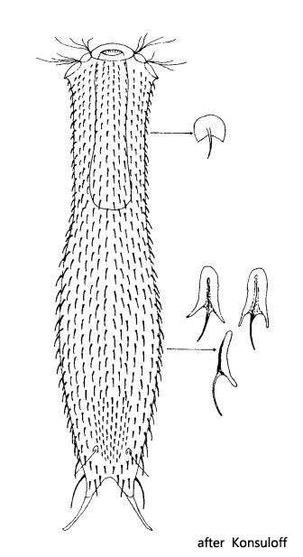
adhesive tubes reaching 3/4 of toe length (16–17 µm)
dorsally 13–17 longitudinal rows of scales with short spines
spines become longer towards posterior end
2–3 pairs of thickened scales at base of toes with long spines
head and neck spines 2–3 µm long
trunk spines 5–7 µm long
at the posterior end a median field of 9–10 longitudinal rows of minute spines
head and neck scales semicircular with a distal incision
trunk scales with a keeled elongate proximal lobe and two distal wings
ventrally 9 longitudinal rows of roundish scales with short spines pharynx cylindrical, terminally slightly swollen
Chaetonotus elegans
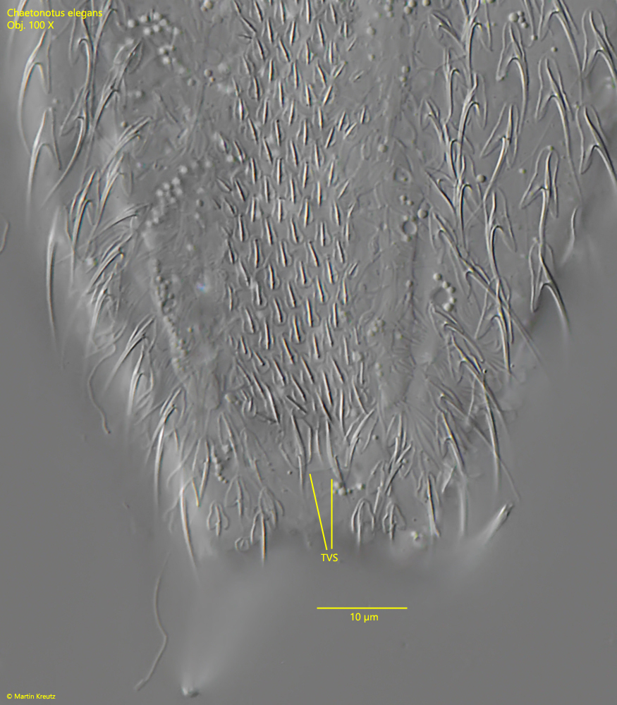
I have found Chaetonotus elegans in November 2021 and in November 2022 in the Simmelried. Both specimens were from the uppermost mud layer. A close examination of the dorsal and ventral scales is necessary to identify this species with certainty. Especially the ventral scales (s. fig. 8) are not easy to document, because the specimens immediately turn on their ventral side under the cover glass. The exact shape and arrangement of the ventral scales has also not been known to my knowledge, as they are only described as “roundish with fine spines”. However, they have a waisted shape and are keeled with very short spines. In addition, there are two diamond-shaped terminal scales just before the base of the toes, which are also keeled, but with a slightly longer spine (s. fig. 8).
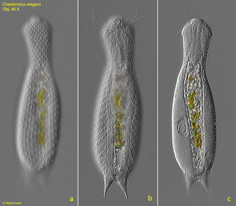
Fig. 1 a-c: Chaetonotus elegans. L = 170 µm. Dorsal view of a freely swimming specimen. Obj. 40 X.
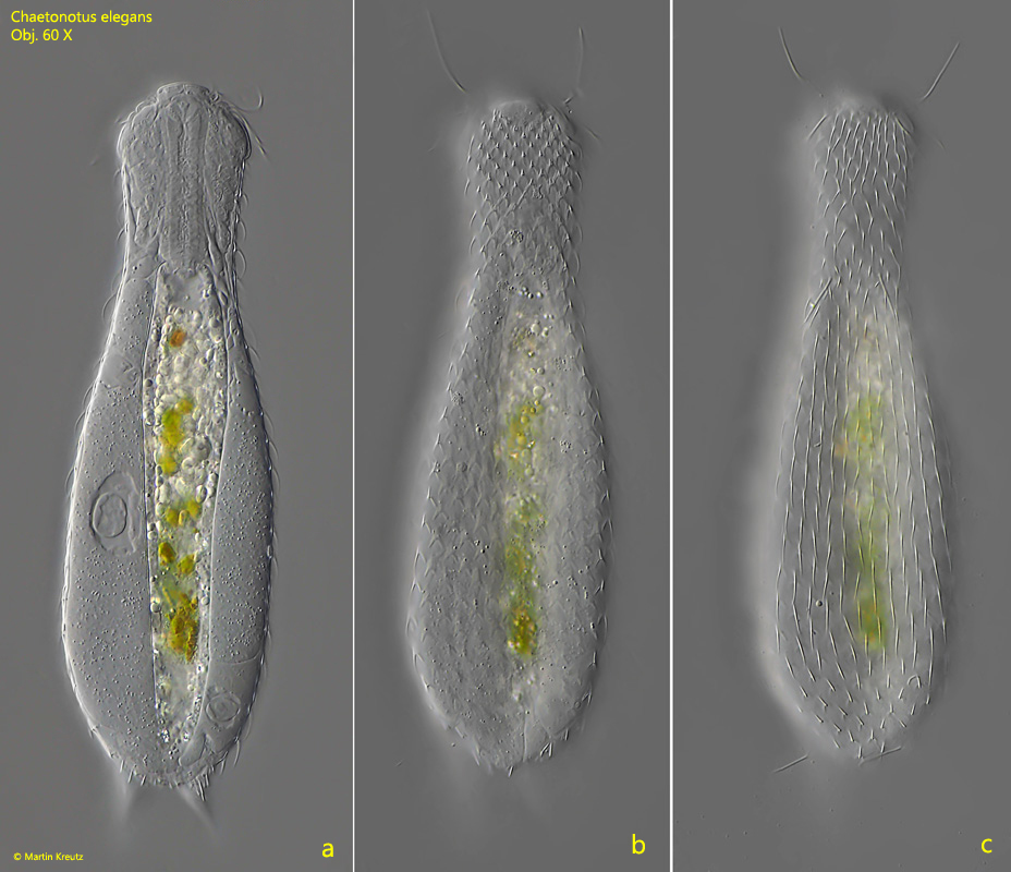
Fig. 2 a-c: Chaetonotus elegans. L = 170 µm. Dorsal view of a slightly squashed specimen. Obj. 60 X.
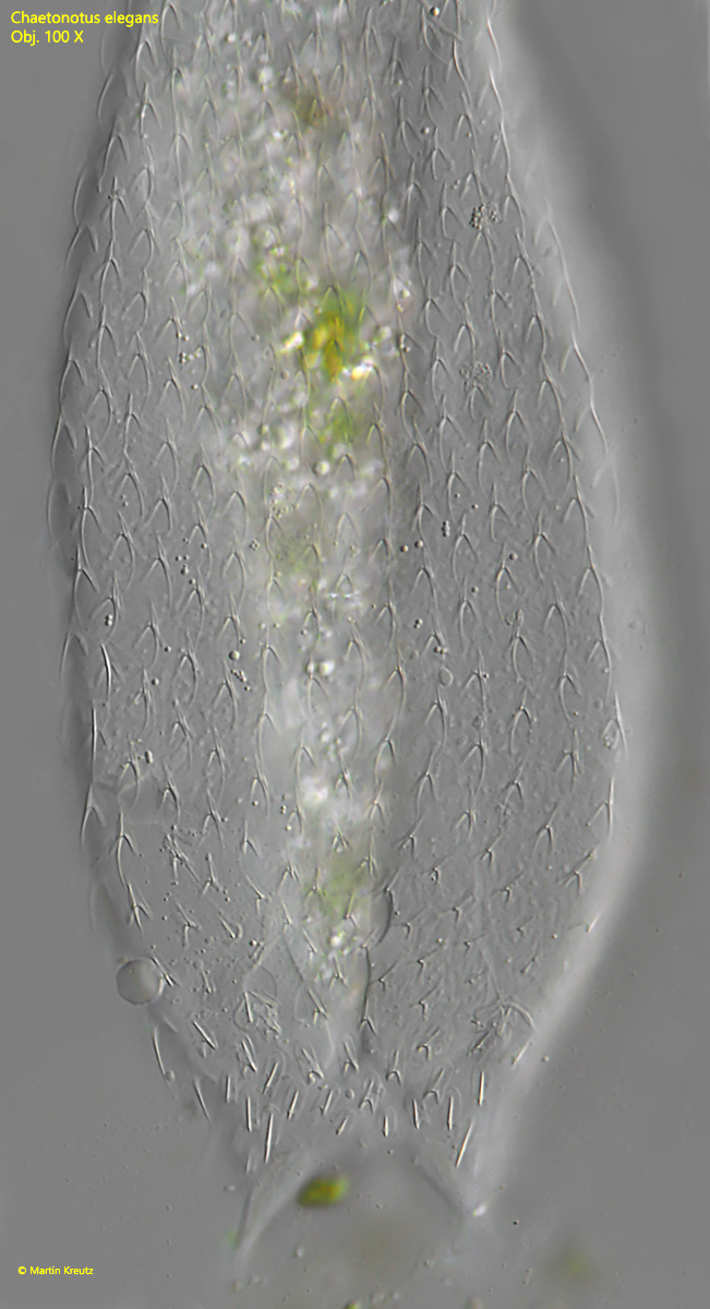
Fig. 3: Chaetonotus elegans. L = 170 µm. Dorsal view of a squashed specimen. Obj. 100 X.
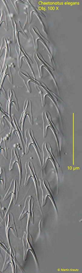
Fig. 4: Chaetonotus elegans. The scales of the neck region in detail. Obj. 100 X.
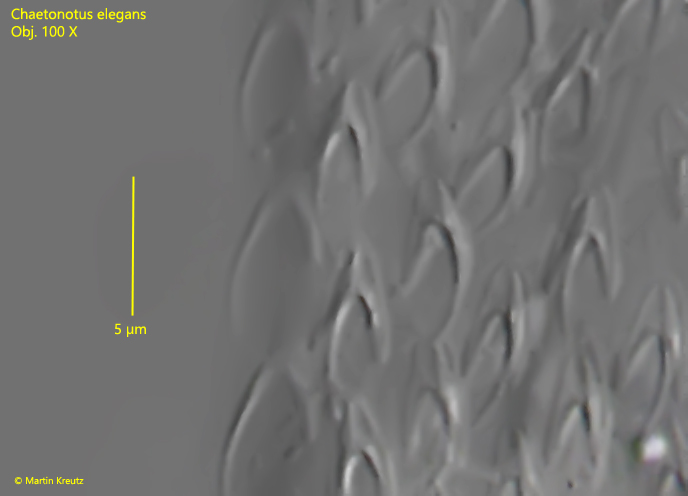
Fig. 5: Chaetonotus elegans. The trilobed scales of the mid-body. Obj. 100 X.
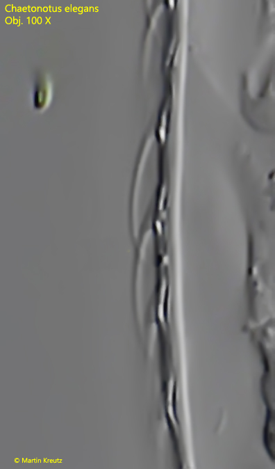
Fig. 6: Chaetonotus elegans. A lateral view of the scales in mid-body. Obj. 100 X.
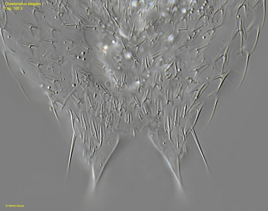
Fig. 7: Chaetonotus elegans. The dorsal scales at the posterior end in detail. Obj. 100 X.
Fig. 8: Chaetonotus elegans. The ventral scales at the posterior end in detail. Note the pair of diamond-shaped terminal ventral scales (TVS). Obj. 100 X.
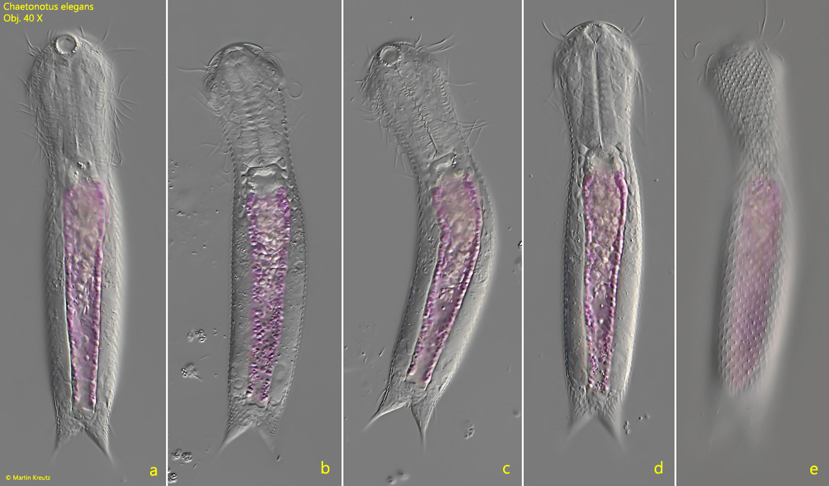
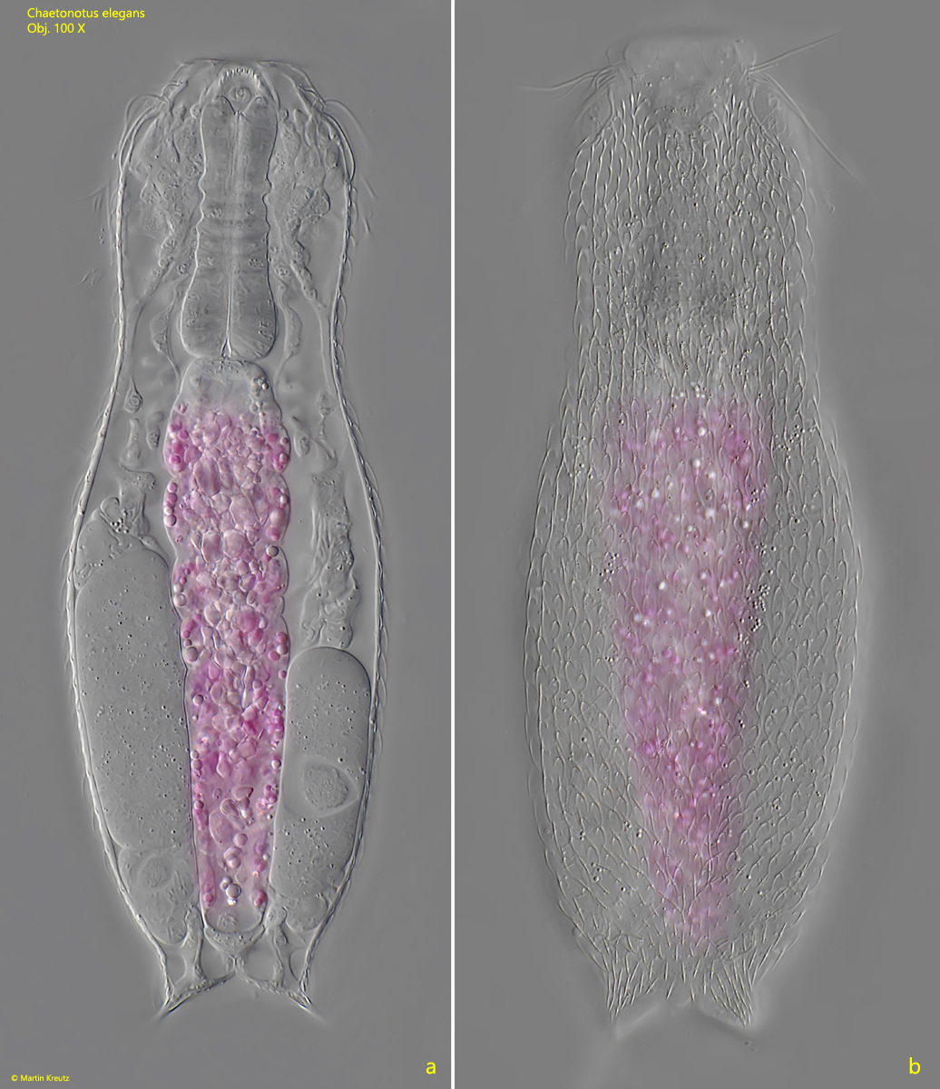
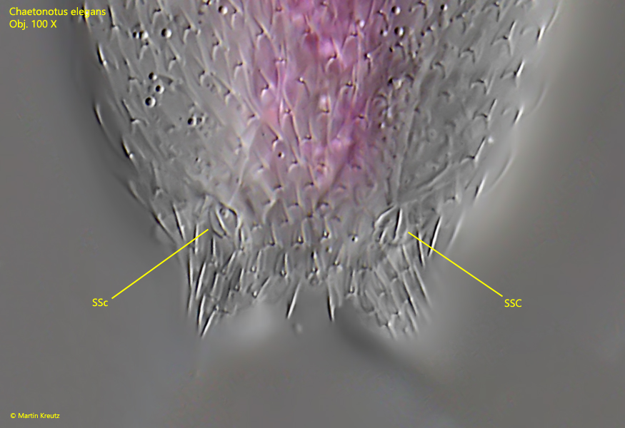
In November 2022, I found a specimen of Chaetonotus elegans in Simmelried with a pink colored intestine (s. figs. 8 a-e and 9 a-b). The color is probably due to phagocytosed rhodobacteria, which were abundant in the sample. In this specimen I could also document the posterior special scales from which the setolae arise (s. fig. 10).
Fig. 8 a-e: Chaetonotus elegans. L = 173 µm. A second freely swimming specimen from dorsal with a pink colored intestine due to phagocytosed rhodobacteria. Obj. 40 X.
Fig. 9 a-b: Chaetonotus elegans. L = 173 µm. Two focal planes of the specimen with the pink intestine from dorsal . Obj. 100 X.
Fig. 10: Chaetonotus elegans. The posterior end of the second specimen in detail. The two special scales (SSC) bearing the setolae are visible. Obj. 100 X.