base of scales trilobed with 6.5–9 µm long simple spines
posteriaor scales keeled with a short spine
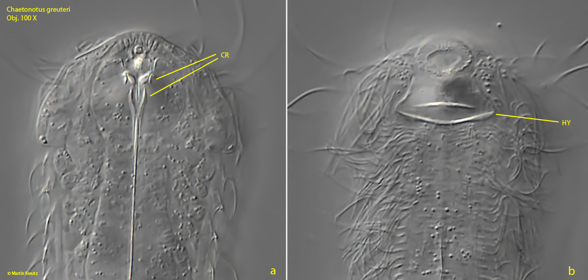
pharynx 30–38 µm long, with conspicuous anterior cuticular reinforcements
adhesive tubes distally pointed
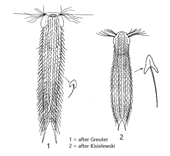
Chaetonotus greuteri
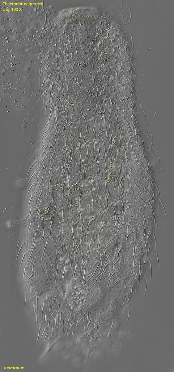
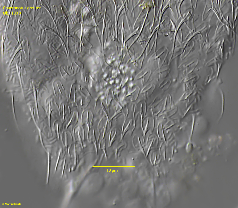
I found several specimens of Chaetonotus greuteri among rotting leaves in Purren Pond. The species seems to be quite variable and there are only few detailed descriptions available, which partly differ from each other. My specimens agreed best with the latest redescription by Kisielewski from 1999 (s. Literature). This also describes for the first time the cuticular reinforcements at the anterior end of the pharynx, whose existence I could also prove (s. fig. 7a). He also mentions for the first time the presence of a hypostomium (s. fig. 7b), which is not mentioned by earlier authors (Greuter, Schwank, s. Literature). In the specimens I examined the shape of the scales in the middle of the body were clearly three-lobed and agreed best with the descriptions and the drawing of Kisielewski. However, supplementing the description by Kisielewski, I was able to demonstrate that the scales abruptly reduce in size at the posterior end and then exhibit a keel with a short spine (s. fig. 6).
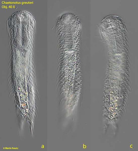
Fig. 1 a-c:Chaetonotus greuteri. L = 150 µm. Dorsal view of a freely swimming specimen. Obj. 40 X.
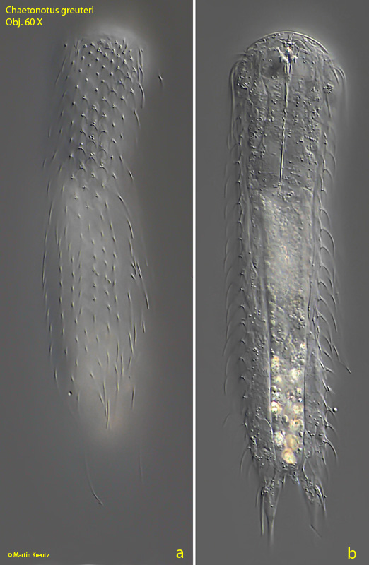
Fig. 2 a-b:Chaetonotus greuteri. L = 150 µm. Dorsal view of the slightly squashed specimen. Obj. 60 X.
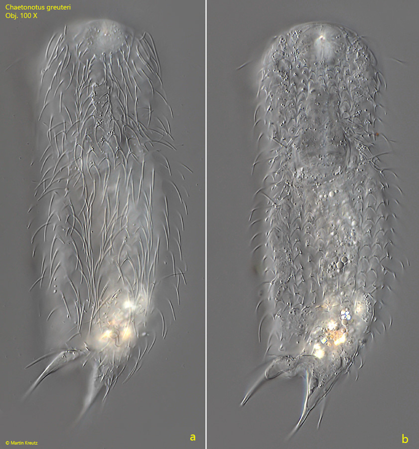
Fig. 3 a-b:Chaetonotus greuteri. L = 150 µm. Dorsal view of the slightly squashed specimen at higher magnification. Obj. 100 X.
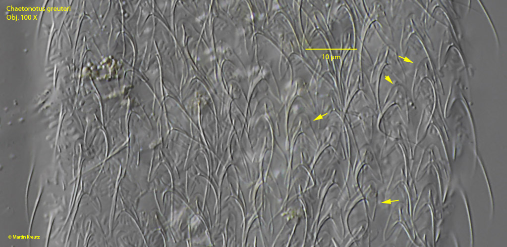
Fig. 4:Chaetonotus greuteri. L = 150 µm. Dorsal view of a strongly squashed specimen to demonstrate the scales in detail. Obj. 100 X.
Fig. 5:Chaetonotus greuteri. L = 150 µm. Detail of fig. 4. The arrows point to scales, whose trilobate shape can be clearly seen. Obj. 100 X.
Fig. 6:Chaetonotus greuteri. L = 150 µm. Detail of fig. 4. The dorsal posterior scales in detail. Obj. 100 X.
Fig. 7 a-b:Chaetonotus greuteri. a) The pharynx with anterior cuticular reinforcements (CR). b) Focus on the ventrally located hypostomium (HY). Obj. 100 X.
Fig. 8:Chaetonotus greuteri. The X-bodies (arrows) in a strongly squashed specimen. Obj. 100 X.
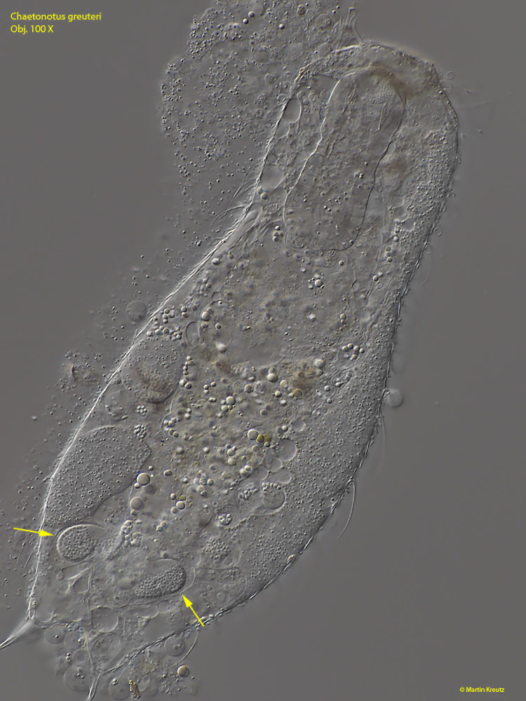
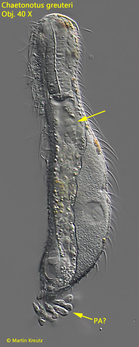
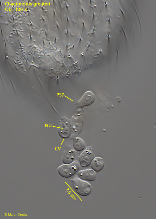
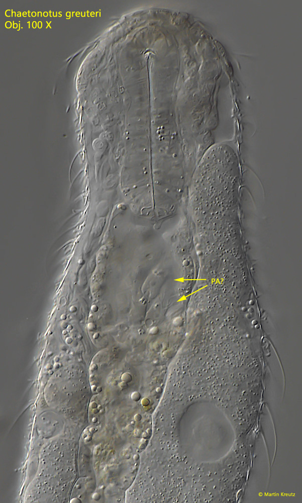
One of the specimens I examined caught my eye by having a cluster of cells trailing behind it. The nature of these cells was difficult to discern in the free-swimming specimen. In the anterior end of the intestine of the specimen, more cells of this type could be recognized (s. fig. 9). In the fixed and squashed specimen, at higher magnification, the trailing cells, which were connected to the specimen in an unknown manner, showed a distinctly amoeboid character. All of the 7–9 µm long cells possessed a nucleus with nucleolus, a contractile vacuole, and also signs of pseudopodia, although I did not see these actively moving (s. fig. 10). In the intestine I could detect the same cells with much longer and branched pseudopodia (s. fig. 11). I therefore believe that this may be a case of parasitism by parasitic amoebae in the intestine of a gastrotrich. To my knowledge, a comparable case of parasitism in gastrotrichs has not yet been described.
Fig. 9:Chaetonotus greuteri. A specimen in lateral view with amoeboid cells in the anterior part of the intestine and a bundle of these cells trainling outside of the body. PA = likely parasitic amoeboid cells. Obj. 40 X.
Fig. 10:Chaetonotus greuteri. The trailing amoeboid cells in detail. All of them had a nucleus (NU) with a central nucleolus, a contractile vacuole (CV) and likely pseudopodia (PS). Obj. 100 X.
Fig. 11:Chaetonotus greuteri. In the intestine of the infested specimen, the amoeboid cells (PA = parasites) can be seen with clearly extended and branched pseudopodia. Obj. 100 X.