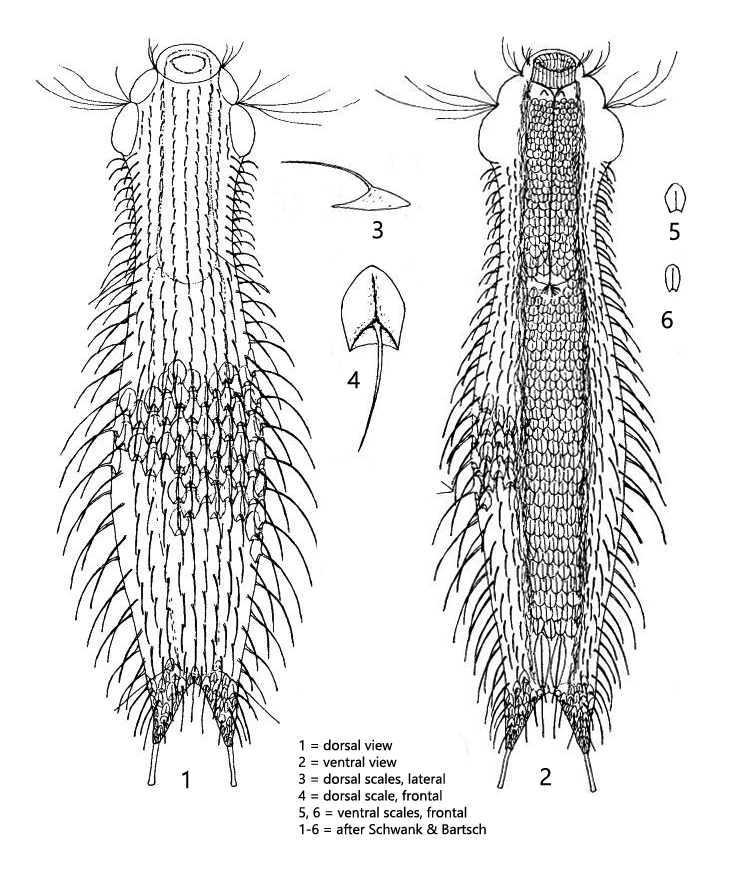
2 pairs of dorsal setolae, posterior on special scales
adhesive tubes 15–19 µm long
furca 27–30 µm long
dorsal scales are pentagonal, about 10 µm long in mid-body, in 11–17 longitudinal rows
dorsal spines arise slightly distal from the scale center
spines curved (10–22 µm long) without a secondary tip
head scales are small (2–3 µm long), roundish, without keel, with short spine
ventrally completely covered with 8–11 longitudinal rows of overlapping, keeled scales with short spines
ventrally two large, elongated terminal scales with keel at posterior end
Chaetonotus laroides
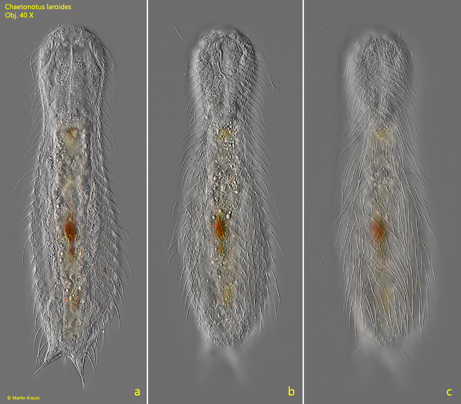
I have found Chaetonotus laroides so far exclusively in the Simmelried between decomposing plant masses but also in the uppermost mud layer. There the species is very common. In my population the specimens were between 280–310 µm long (inclusive toes) and thus clearly larger than indicated by Schwank and Bartsch, 1990 (s. Literature). The specimens are conspicuous by their densely standing spines, which are clearly curved (s. figs. 1 a-c and 2 a-b). Often the specimens carry an egg (s. fig. 2 a-b). The dorsal scales are pentagonal in the middle of the body (s. figs. 5 and 6), whereas the distal margin is very thin and difficult to recognize (s. fig. 6). Small magnifications may therefore give the impression that the dorsal scales are trilobate. In lateral view, the anterior margin of the scales appears hooked (s. fig. 7). One can assume that this structure serves to hook the scales together. At the posterior end I could always see 3 straight spines between the toes (s. fig. 9). These are not mentioned by Schwank and Bartsch, but seem typical to me.
Fig. 1 a-c:Chaetonotus laroides. L = 294 µm. Three focal planes of a freely swimming specimen. Obj. 40 X.
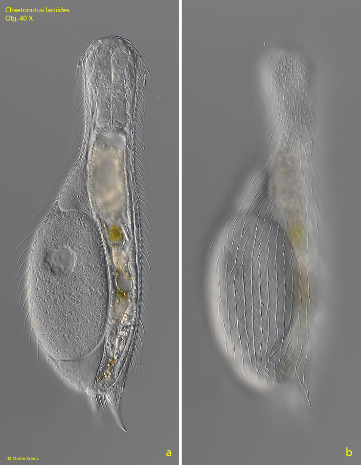
Fig. 2 a-b:Chaetonotus laroides. L = 310 µm. A second specimen with an egg. Obj. 40 X.
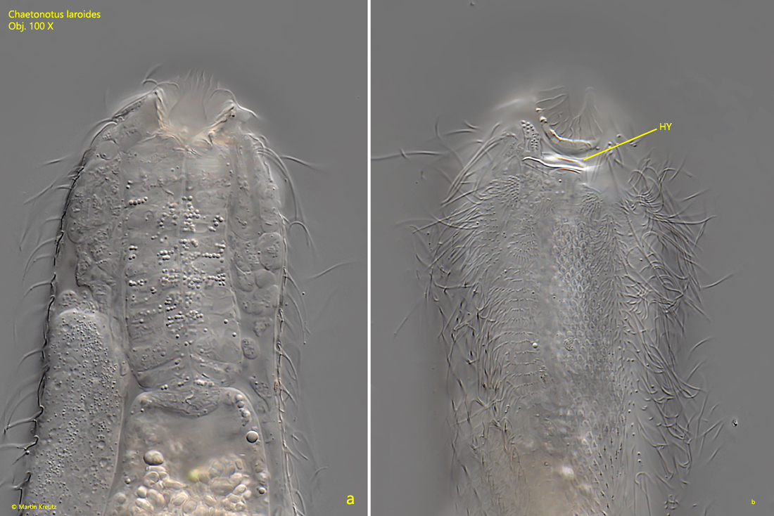
Fig. 3 a-b:Chaetonotus laroides. Two focal planes of the head from the ventral side. HY = hypostomium. Obj. 100 X.
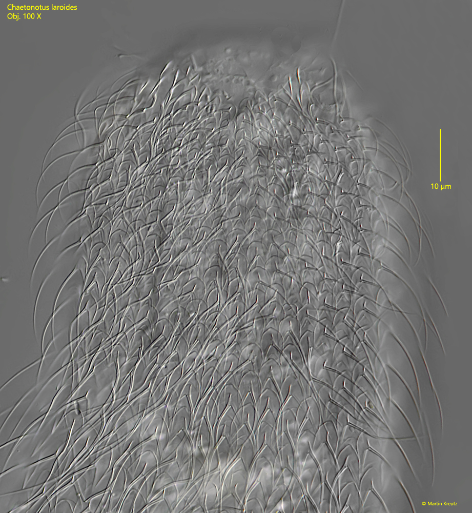
Fig. 4:Chaetonotus laroides. The dorsal scales in the head region. Obj. 100 X.
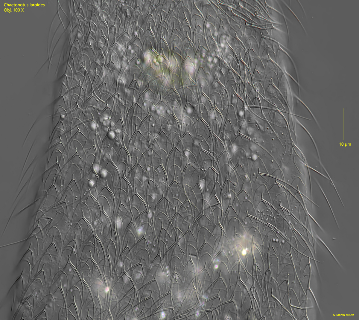
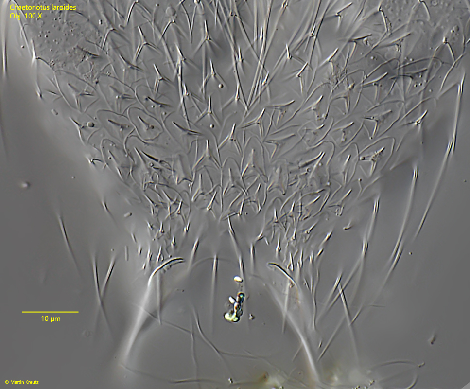
Fig. 5:Chaetonotus laroides. The dorsal scales of the mid-body. Obj. 100 X.
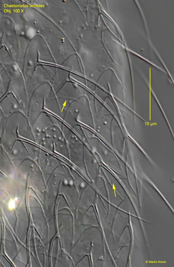
Fig. 6:Chaetonotus laroides. The almost pentagonal dorsal scales in detail. The distal margin of the scales (arrows) is thin and hard to see. Obj. 100 X.
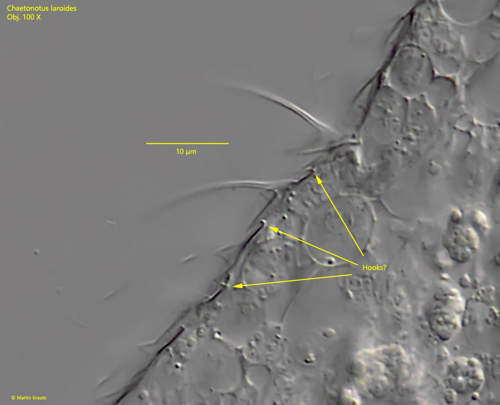
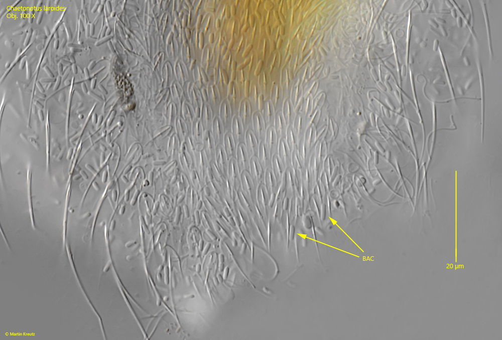
Fig. 7:Chaetonotus laroides. In lateral view of the dorsal scales, it looks as if the anterior margin is folded over, giving a hook-like structure (arrows). Presumably, this causes the scales to be hooked into each other. Obj. 100 X.
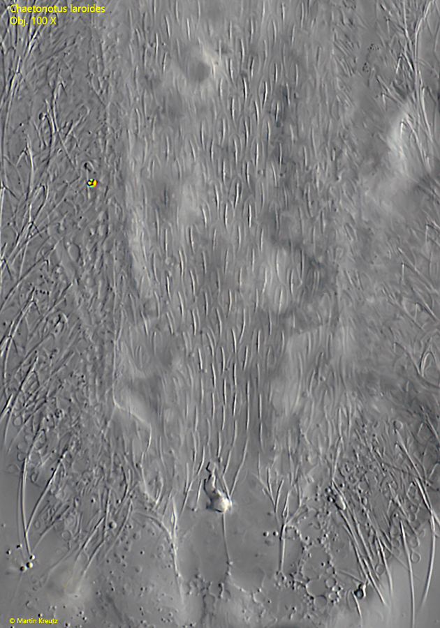
Fig. 8:Chaetonotus laroides. The dorsal scales at the posterior end in detail. Obj. 100 X.
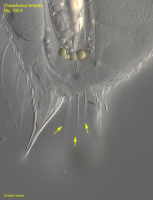
Fig. 9:Chaetonotus laroides. At the posterior end three spines between the toes are visible (arrows). Obj. 100 X.
Fig. 10:Chaetonotus laroides. The ventral scales at the posterior end focussed from the dorsal side. Obj. 100 X.
Fig. 11:Chaetonotus laroides. The ventral scales at the posterior end of a second specimen. Obj. 100 X.