dorsal scales pouchlike with a pattern of transverse and longitudinal stripes
At the end the scales elongate to a spine with a secondary tip
dorsal scales arranged in longitudinal rows, not overlapping
ventral scales keeled, trilobed basal plate
cephalion broad, pleurae reduced
two pairs of tactile ciliary tufts
hypostomiun present
pharynx large, about 60 µm long
adhesive tubes slightly curved
Chaetonotus robustus
Chaetonotus robustus appears to be a comparatively rare gastrotrich. So far only findings from Poland and the USA are known. I found Chaetonotus robustus so far exclusively in the Simmelried. The first finding is from 2004, after that I found it again and again in larger intervals. With increasing siltation of the Simmelried the findings seem to accumulate. It is found exclusively in the upper mud layer, especially where larger plant masses are rotting. Due to its size of more than 500 µm it is immediately noticeable even at small magnification. The locomotion is a slow gliding and burrowing. Confusion is also impossible due to its uniquely shaped, pouch-like scales.
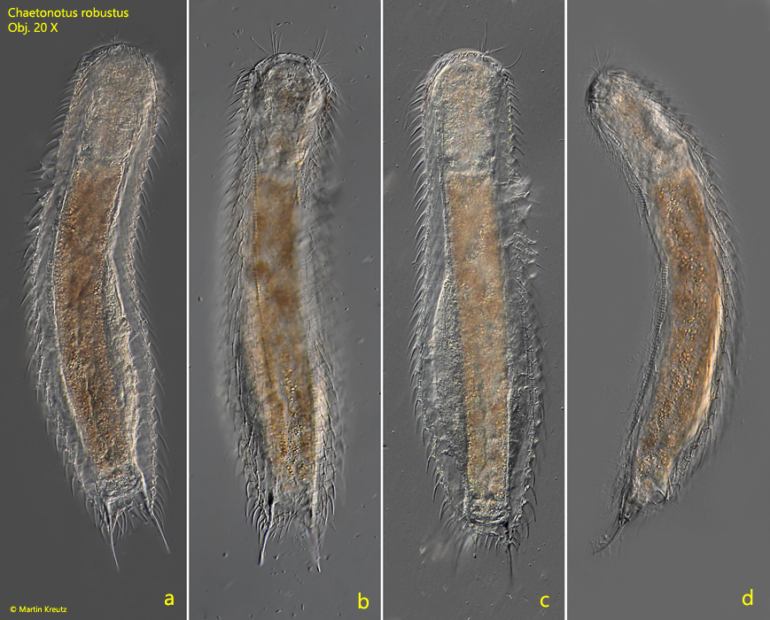
Fig. 1 a-d:Chaetonotus robustus. L = 530 µm. A freely swimming specimen. a, b) ventral view, c) dorsal view, d) lateral view from right. Obj. 40 X.
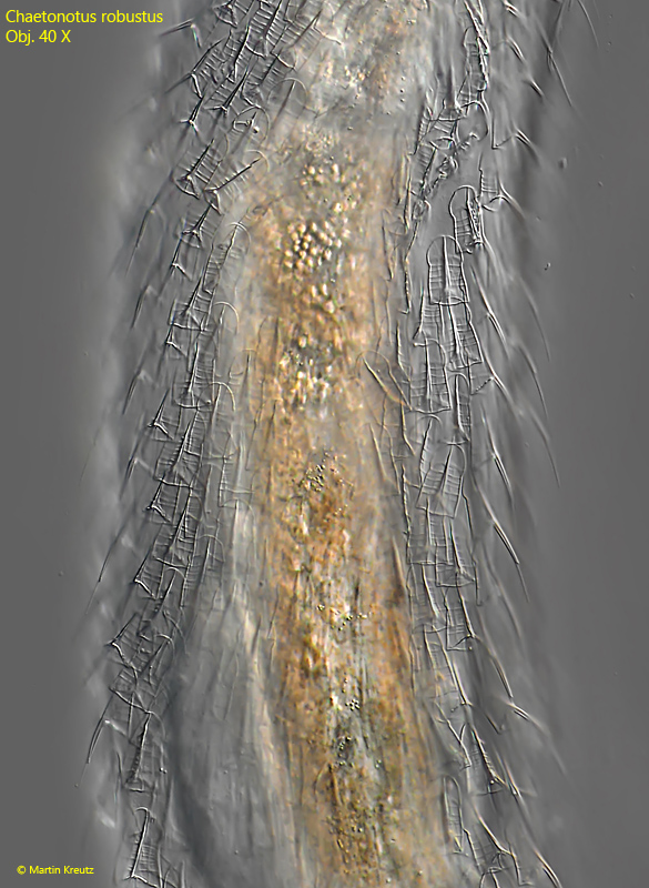
At medium magnification and in dorsal view, it almost appears as if the specimens are draped with glassy bells (s. fig. 2).
Fig. 2:Chaetonotus robustus. L = 530 µm. View of the dorsal scales of a slightly squashed specimen. Obj. 40 X.
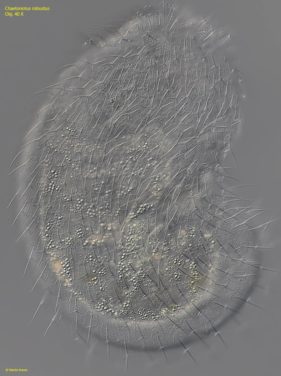
Fig. 3:Chaetonotus robustus. Dorsal view of the scales of a squashed and contracted specimen. Obj. 40 X.
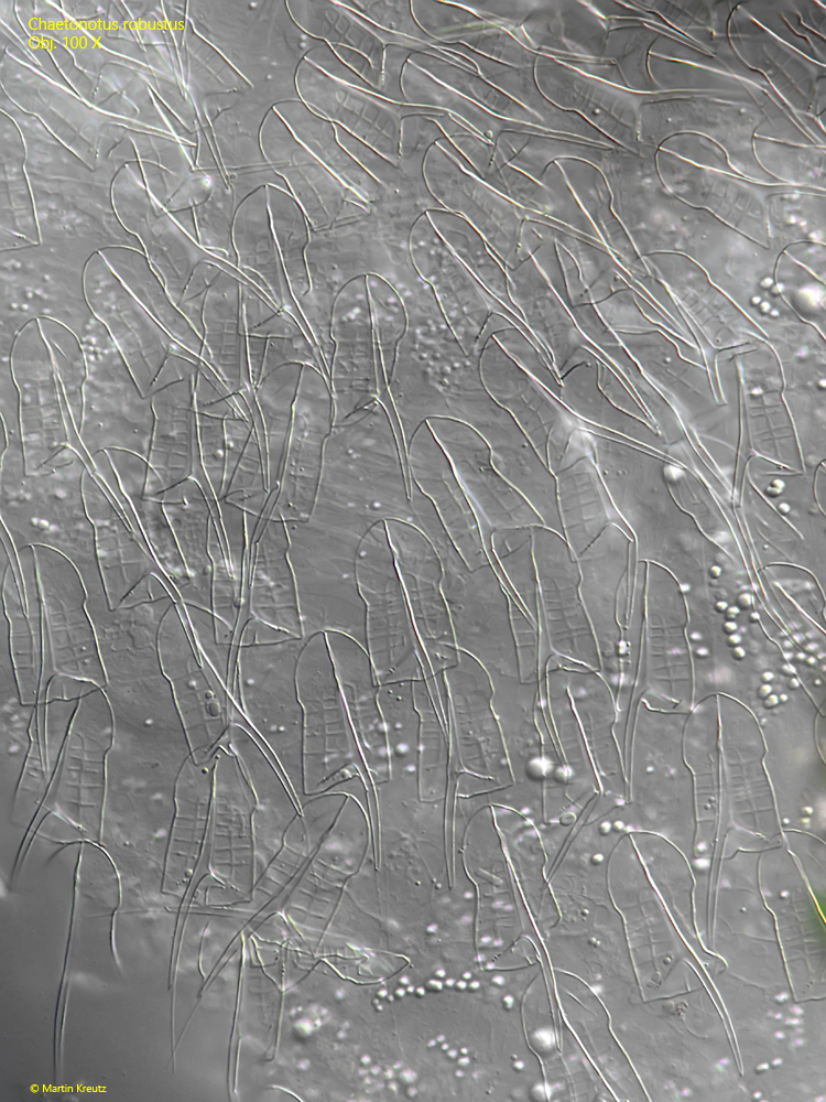
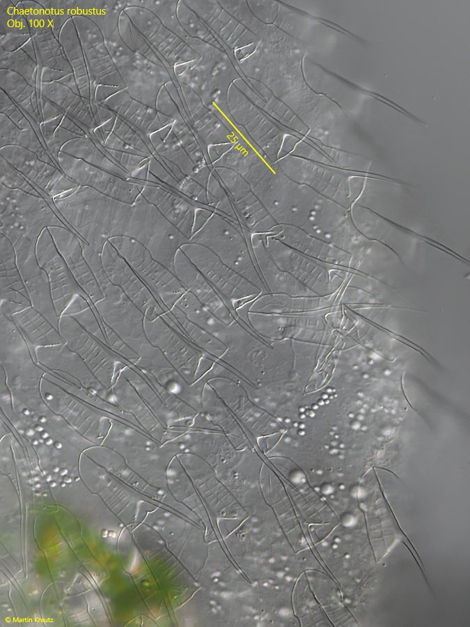
Fig. 4:Chaetonotus robustus. The dorsal scales in mid-body of a strongly squashed specimen. Obj. 100 X.
Fig. 5:Chaetonotus robustus. The dorsal scales on the edge of the body of a strongly squashed specimen. Obj. 100 X.
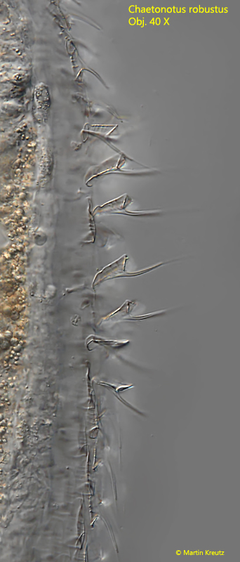
Fig. 6:Chaetonotus robustus. Coverslip pressure sometimes causes the lateral scales to be dislodged from their original position. Obj. 40 X.
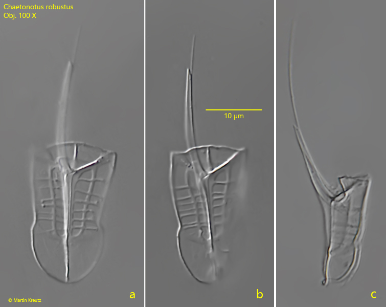
Fig. 7 a-c:Chaetonotus robustus. The detached dorsal scales in detail. Note the lattice-like pattern and the elongated distal spine. The total length of the scales in mid-body (including spine) is about 50 µm. a, b) frontal view, c) lateral view. Obj. 100 X.
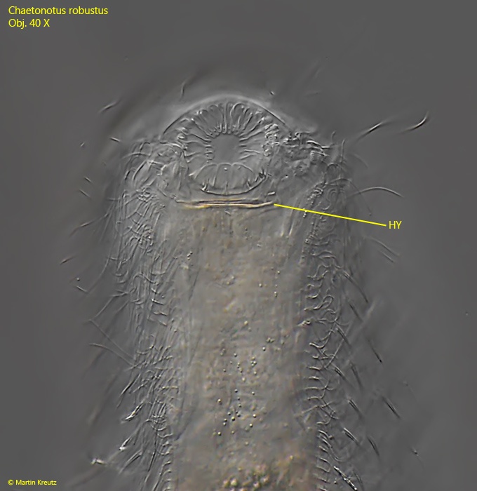
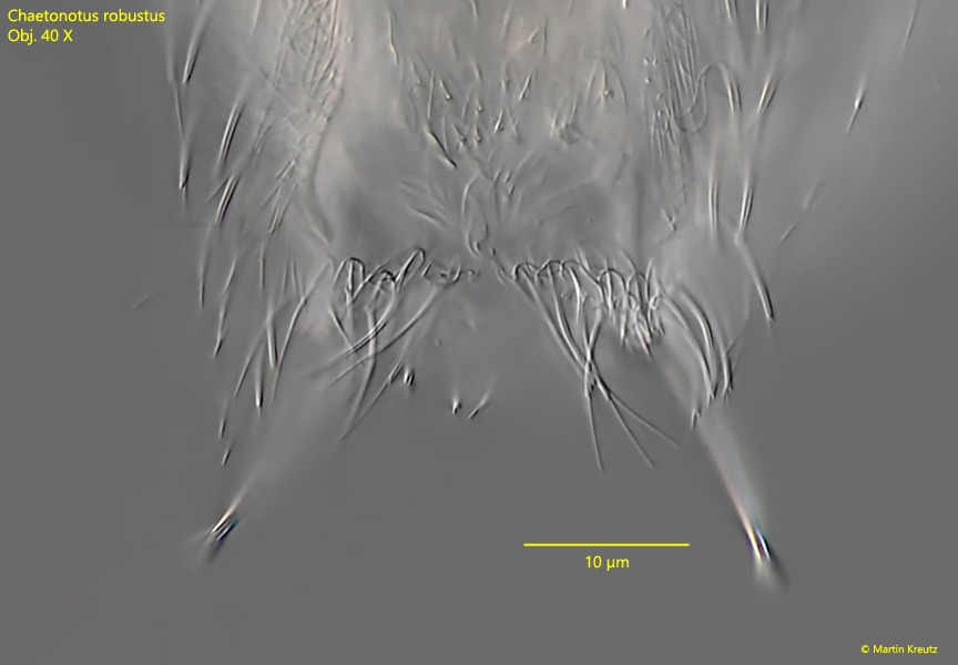
I could also examine specimens in ventral view. I found that a hypostomium is present, which was not described by earlier authors (s. fig. 8). The shape of the ventral scales differs considerably from that of the dorsal scales. At the level of the pharynx they are arrow-shaped with a keel (s. fig. 9) and in the middle of the body they are three-lobed, also with a keel and a short spine (s. fig. 10).
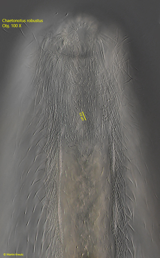
Fig. 8:Chaetonotus robustus. L = 530 µm. Ventral view of the mouth opening surrounded by overlapping plates. HY = hypostomium. Obj. 40 X.
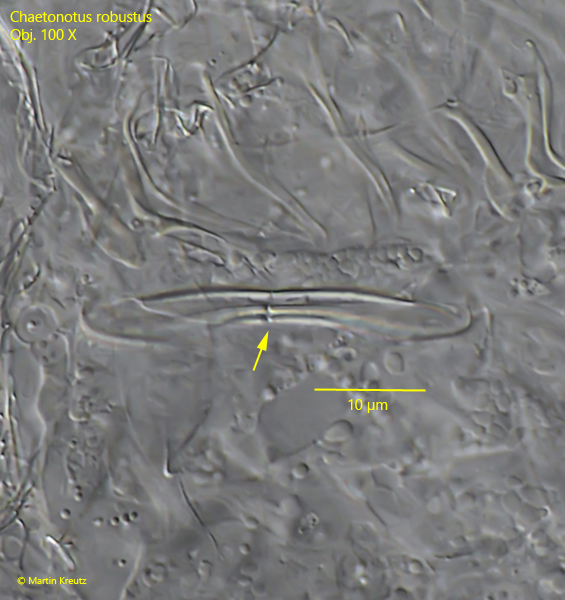
Fig. 9:Chaetonotus robustus. Detail of the hypostomium in a strongly squashed specimen. A characteristic gap is present in the middle of the hypostomium (arrow). Obj. 100 X.
Fig. 10:Chaetonotus robustus. L = 530 µm. Ventral view of the scales at the level of the pharynx. Obj. 100 X.
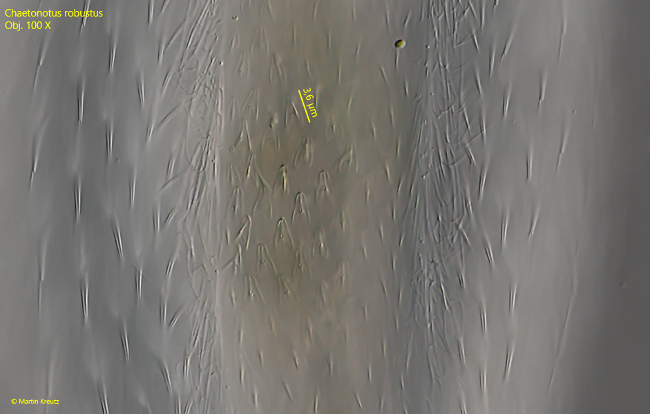
Fig. 11:Chaetonotus robustus. L = 530 µm. Ventral view of the scales in mid-body. Obj. 100 X.
Fig. 12:Chaetonotus robustus. L = 530 µm. Ventral view of the scales at the posterior end. Obj. 100 X.
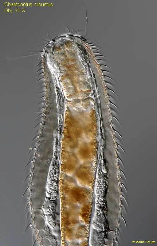
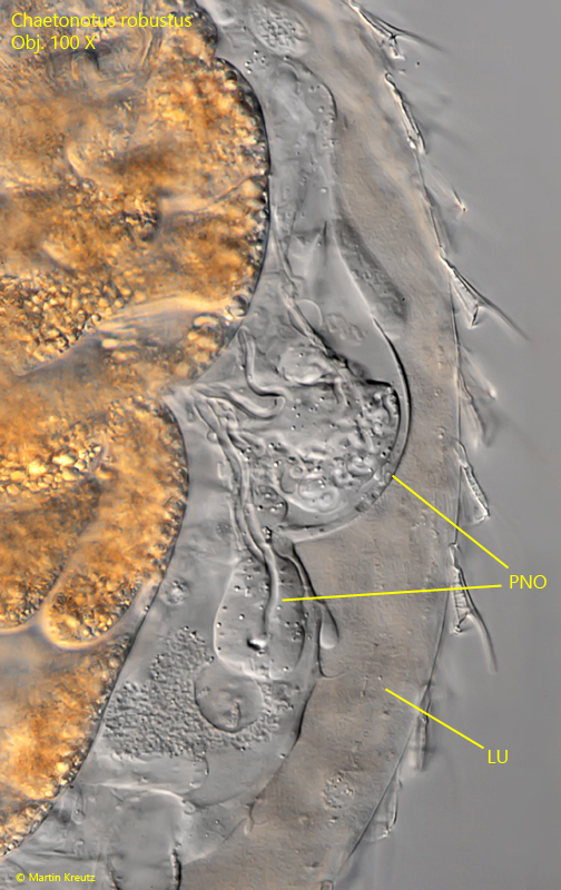
Another characteristic feature of Chaetonotus robustus is a lumen (i.e. a spacious body cavity unlike in most other gastrotrichs) separating the internal organs from the cuticle with scales (s. figs. 12 and 13). I could observe this in several specimens.
Fig. 13:Chaetonotus robustus. The cuticle with the scales is set apart from the intestine and inner organs by a lumen. Obj. 20 X.
Fig. 14:Chaetonotus robustus. Focus on the protonephridial organ and the lumen between the organs and the cuticle. LU = lumen, PNO = protonephridial organ. Obj. 100 X.
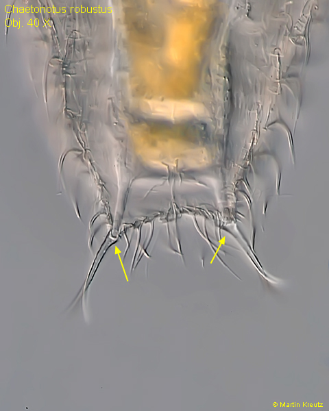
Fig. 15:Chaetonotus robustus. The adhesive tubes are slightly curved. At the origin they show a concave indentation (arrows). Obj. 40 X.
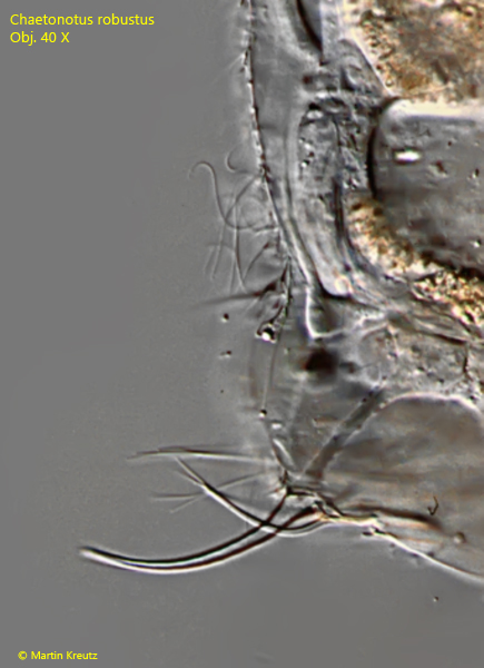
Fig. 16:Chaetonotus robustus. Detail of the adhesive tubes in a squashed specimen. Obj. 40 X.
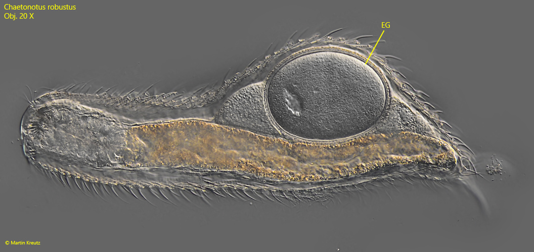
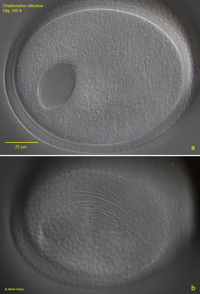
I have found so far only one specimen with one egg (s. fig. 16). I took the opportunity to examine the egg at high magnifications. The egg shell has a very faint hexagonal pattern that is difficult to see in DIC (s. fig. 17). It is not impossible that this hexagonal pattern intensifies just before egg laying or after egg laying.
Fig. 17:Chaetonotus robustus. L = 550 µm. Lateral view of a specimen with an egg (EG). Obj. 20 X.
Fig. 18 a-b:Chaetonotus robustus. Two focal planes of the egg. The shell of the egg reveals a hexagonal pattern (b). Obj. 100 X.
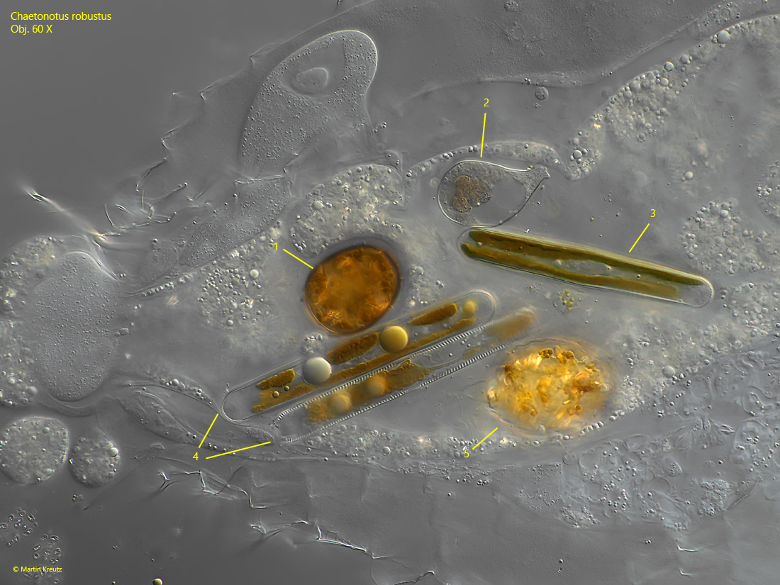
Fig. 19:Chaetonotus robustus. The content of the intestine in a squashed specimen. Some of the undigested prey organisms can be identified as Trachelomonas spec. (1), Notosolenus spec. (2), Pinnularia spec. (3, 4) as well as an unidentified organism (5). Obj. 60 X.