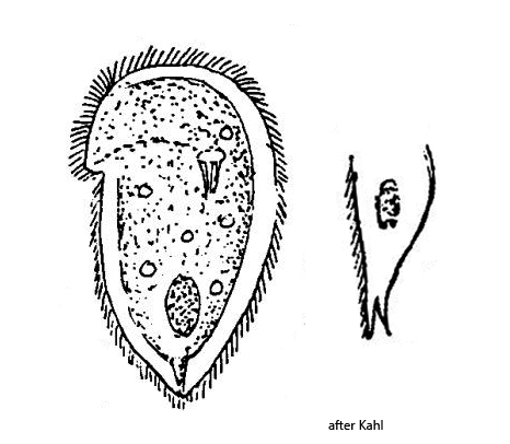
a curved dorsal brush in the center of the anterior fourth
ventral ciliation of Chilodonella type
several contractile vacuoles
oval macronucleus in posterior half
one spherical micronucleus adjacent to macronucleus
Chilodonella caudata
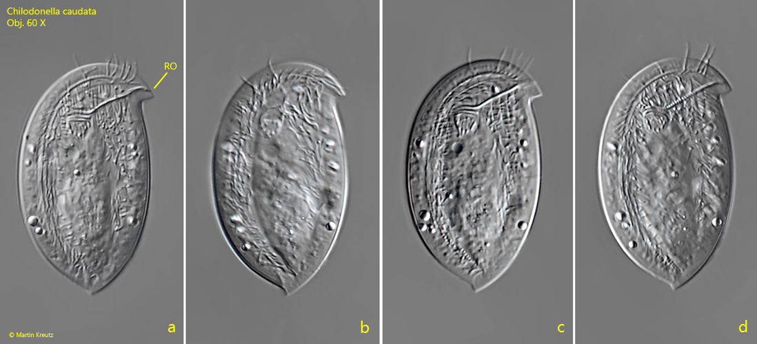
I regularly find Chilodonella caudata in samples with decomposing plant masses and on the floating coverslip. The species can be easily recognized by two features. Firstly, the rostrum on the left protrudes at an almost right angle (s. fig. 1 a-d) and there is a conspicuous spine located on the posterior end of the dorsal side (s. figs. 4 and 5 a-c).
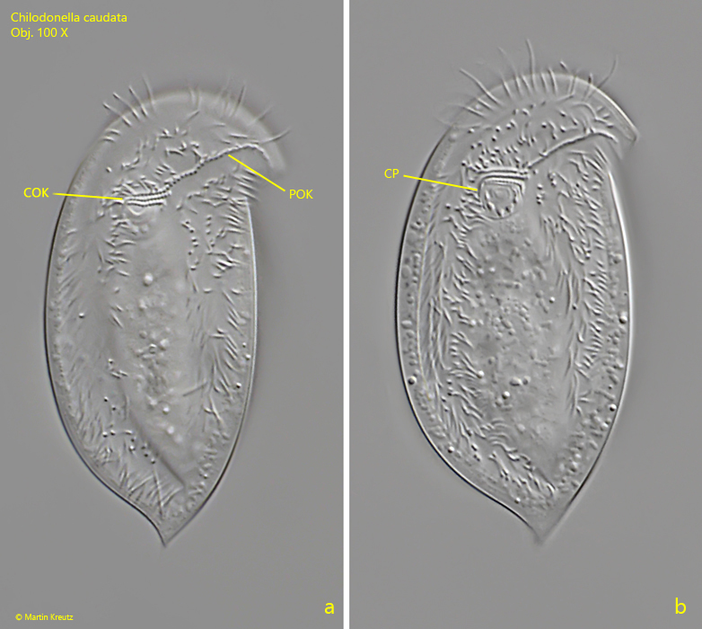
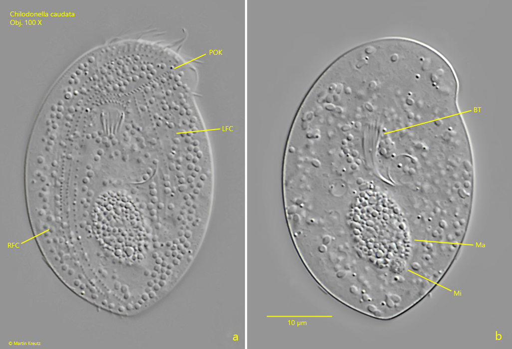
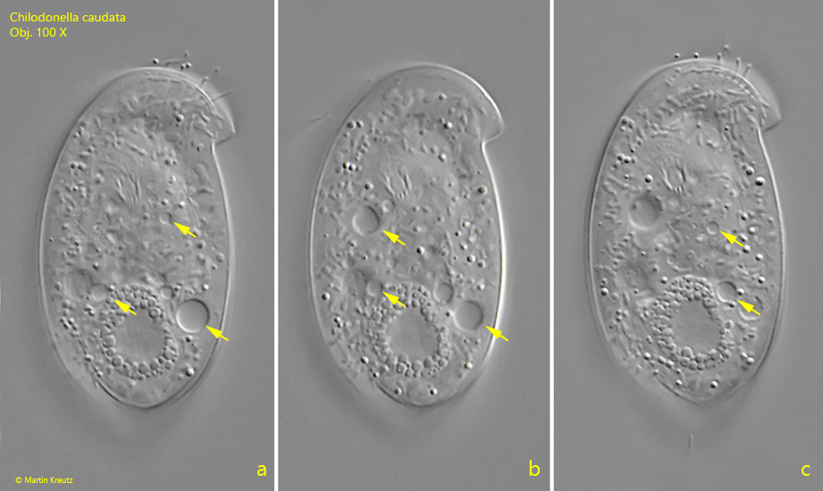
Chilodonella caudata has only rarely been described so far. Kahl reports that the ventral side has not yet been examined. I therefore assume that Kahl did not find Chilodonella caudata himself, but took over the description from Stokes. I was able to examine the ventral side in detail in a squashed specimen (s.e figs. 2 a-b and 3 a). It is characteristic of the genus Chilodonella with a continuous pre-oral kinety, three circumoral kineties and a left and right field of cilia framing a naked midfield. The specimens in my population were between 36–50 µm long. There are several contractile vacuoles present (at least 5, s. fig. 8 a-c) as described by Kahl.
Fig. 1 a-d:Chilodonella caudata. L = 47 µm. A freely gliding specimen from ventral. RO = rostrum. Obj. 60 X.
Fig. 2 a-b:Chilodonella caudata. L = 49 µm. Two focal planes from the ventral side. Note the pre-oral kinety (POK) and the circumoral kineties (COK). CP = cytopharynx. Obj. 100 X.
Fig. 3 a-b:Chilodonella caudata. Two focal planes from the ventral side of a squashed specimen. The ciliation of the ventral side is divided in a left field of cilia (LFC) and a right field of cilia (RFC) while the middle is naked. BT = basket of trichites, Ma = macronucleus, Mi = micronucleus, POK = pre-oral kinety. Obj. 100 X.
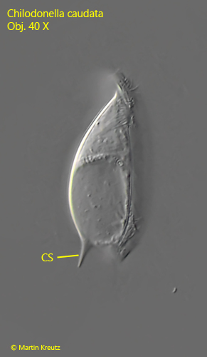
Fig. 4:Chilodonella caudata. Lateral view from the right side. Note the distinct caudal spine (CS) on the dorsal side. Obj. 100 X.
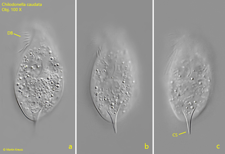
Fig. 5 a-c:Chilodonella caudata. L = 37 µm. Dorsal view of a freely gliding specimen with the distinct caudal spine (CS). DB = dorsal brush. Obj. 100 X.
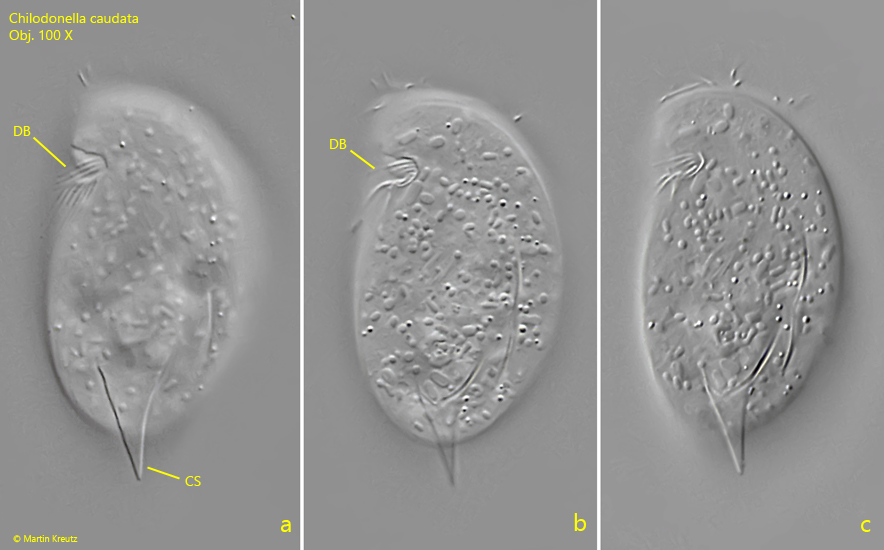
Fig. 6 a-c:Chilodonella caudata. L = 37 µm. The slightly squashes specimen as shown in fig. 5 a-c with focal plane on the dorsal brush (DB). The dorsal brush is located in a depression of the pellicle. Obj. 100 X.
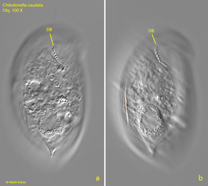
Fig. 7 a-b:Chilodonella caudata. L = 49 µm. Focal plane on the dorsal brush (DB) from the ventral side. Obj. 100 X.
Fig. 8 a-c:Chilodonella caudata. L = 44 µm. Ventral view with focal plane on the contractile vacuoles. In this specimen five contractile vacuole (arrows) are visible. Obj. 100 X.