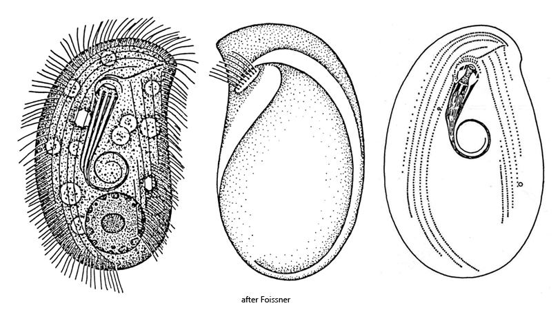
body broadly ellipsoid, rounded anteriorly and posteriorly
dorso-ventrally flattened
rounded or slightly pointed rostrum at anterior third of left side
length 25–70 µm, mostly about 45 µm
ventral side flat, dorsal side convex
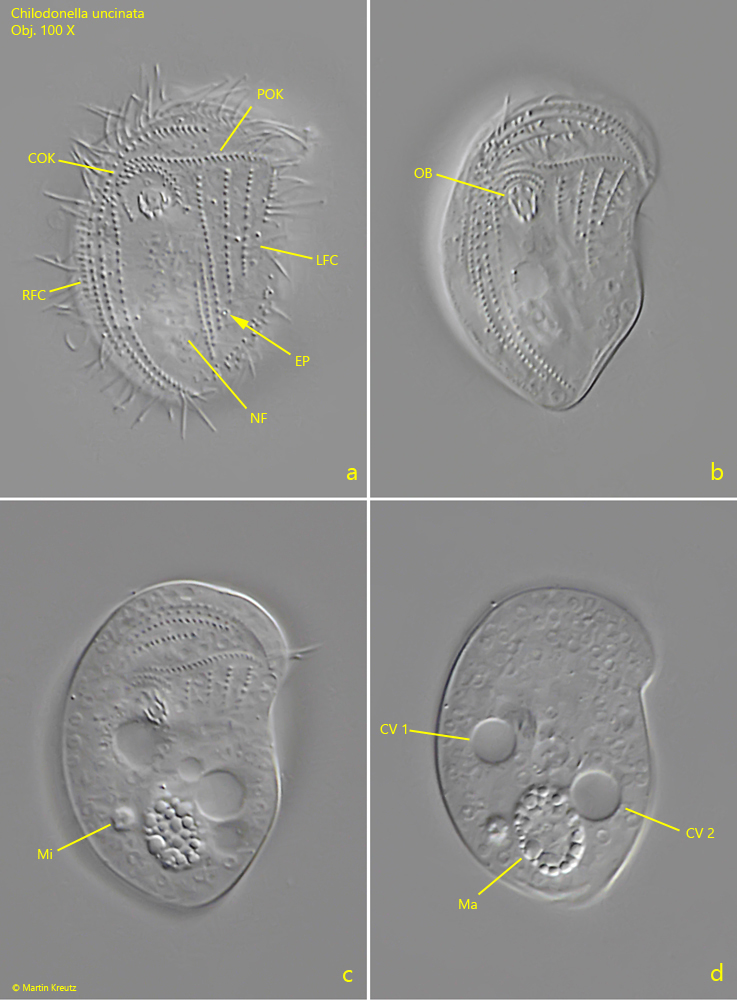
oral basked consisting of (9)–10–(11) rods
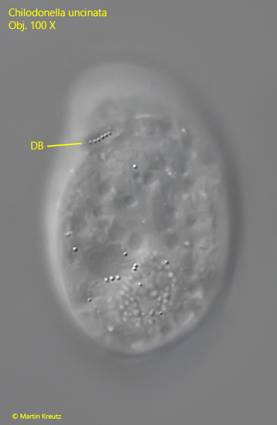
dorsal brush a single row of cilia, subapically near the anterior left margin
two contractile vacuoles in diagonal position, the anterior one on the right side
macronucleus spherical to ellipsoid near posterior end, with many chromatin clods
one spherical micronucleus adjacent to macronucleus
ventrally two fields of cilia separated by a naked field in the middle
two curved circumoral kineties above oral basked
a continuous pre-oral kinety above the circumoral kineties
Chilodonella uncinata
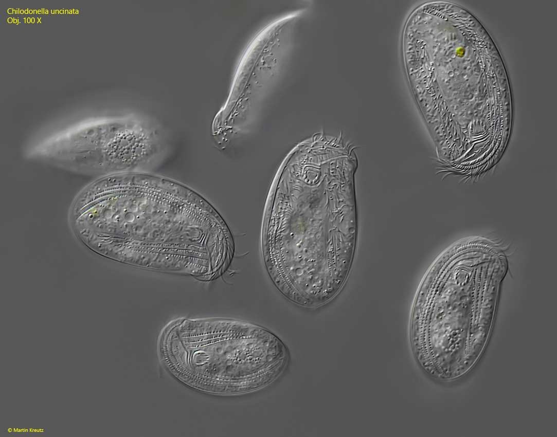
Chilodonella uncinata is one of the most common cyrtophorid ciliates and I find it in practically all of my sampling sites. The specimens collect at the surface in the sample containers where they slide along the water film with their ventral side and feeding on bacteria. Chilodonella uncinata can be observed particularly well on the floating coverslip, where this ciliate regularly colonizes.
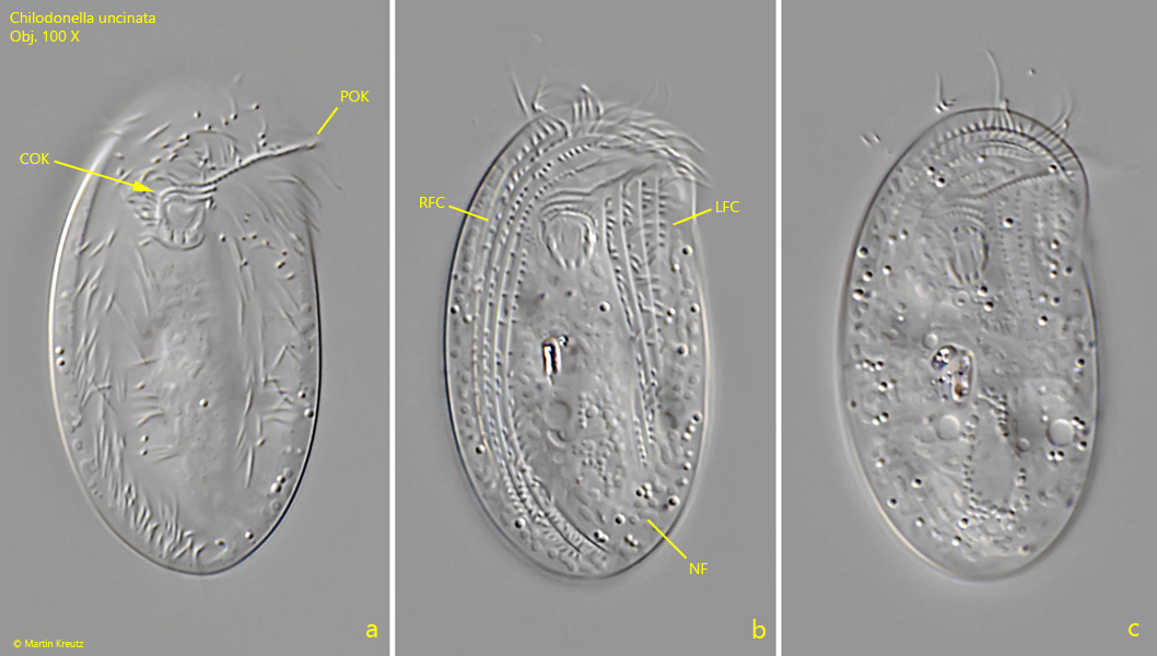
The identification of Chilodonella uncinata is mainly based on the ciliature of the ventral side and the shape and position of the dorsal brush. In Chilodonella uncinata, the dorsal brush is a short row of cilia that is positioned subapically, i.e. does not protrude beyond the anterior margin (s. figs. 3a and 4). Cyrtophorid ciliates with an apical dorsal brush that protrudes beyond the anterior margin belong to the genus Odontochlamys.
On the ventral side of Chilodonella there are three rows of cilia above the mouth opening. The almost semicircular rows of cilia directly adjacent to the mouth opening are known as the circumoral cilia (s. figs. 5 a and 6 b). Above these two rows of circumoral cilia is the pre-oral cilia row located (s. figs. 2 a, 5 a and 6 b), which runs from the mouth opening to the right margin. The pre-oral kinety is continuous and not interrupted in the genus Chilodonella. If it is interrupted like a staircase, then it belongs to the genus Pseudochilodonopsis.
In the middle of the ventral side, Chilodonella uncinata has a well-defined naked field without cilia (s. figs, 2 b, 5 a and 6 b). To the left and right of this naked field are parallel rows of cilia (s. figs. 2 b, 5a and 6 b). If the naked field in the middle is only small and difficult to recognize, then it is the genus Thigmogaster.
Fig. 1:Chilodonella uncinata. L = 38–50 µm. An accumulation of several specimens gliding with their ventral sides along the coverslip. Obj. 100 X.
Fig. 2 a-c:Chilodonella uncinata. L = 40 µm. Different focal planes of a slightly squashed specimen from ventral. Note the continuous pre-oral kinety (POK) and the naked field (NF) between the right and left field of cilia (RFC, LFC). Obj. 100 X.
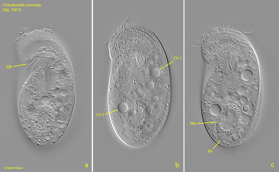
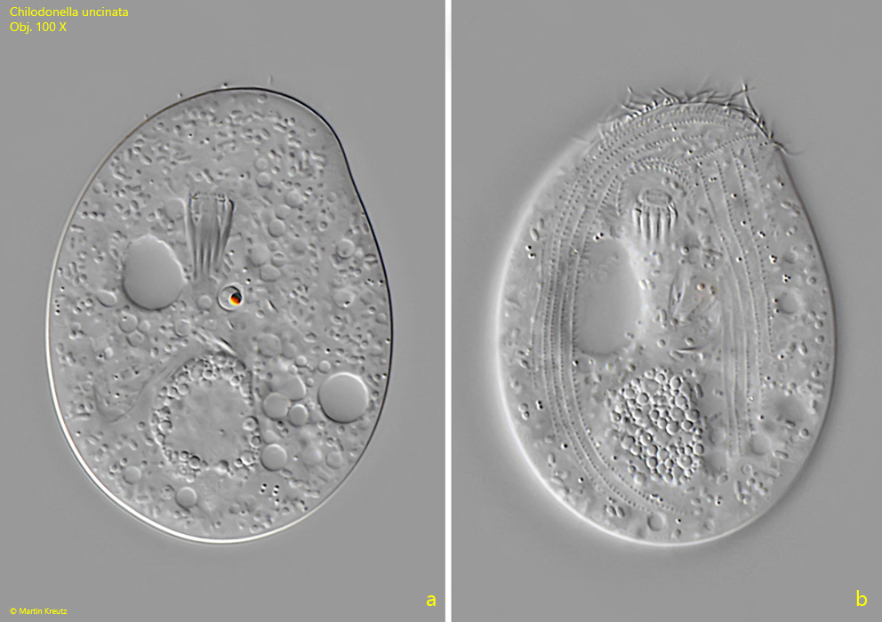
Fig. 3 a-c:Chilodonella uncinata. L = 45 µm. Different focal planes of a slightly squashed specimen from dorsal. Note the dorsal brush (DB), consisting of a short row of cilia and the two contractile vacuoles (CV 1, CV 2) which are arranged diagonally. Ma = macronucleus, Mi = micronucleus. Obj. 100 X.
Fig. 4:Chilodonella uncinata. The dorsal brush (DB) in a squashed specimen from dorsal. Obj. 100 X.
Fig. 5 a-d:Chilodonella uncinata. L = 30 µm. Different focal planes of a squashed specimen from ventral. CV 1, CV 2 = contractile vacuoles, COK = circumoral kineties, EP = excretion porus, LFC = left field of cilia, Ma = macronucleus, Mi = micronucleus, NF = naked field, OB = oral basket, RFC = right field of cilia. Obj. 100 X.
Fig. 6 a-b:Chilodonella uncinata. Two focal planes of a second, squashed specimen from ventral. Obj. 100 X.
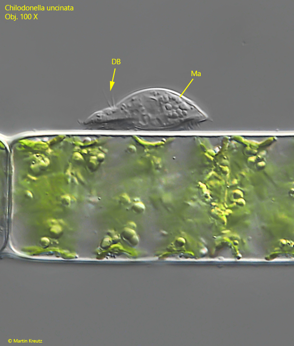
Fig. 7:Chilodonella uncinata. L = 31 µm. Lateral view of a specimen gliding on an algae filament. DB = dorsal brush, Ma = macronucleus. Obj. 60 X.
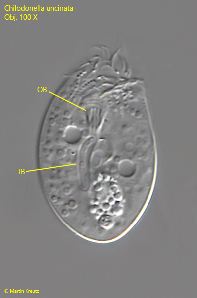
Fig. 8:Chilodonella uncinata. L = 33 µm. A slightly squashed specimen from ventral with an ingested bacterium (IB) in an elongated food vacuole. OB = oral basket. Obj. 100 X.
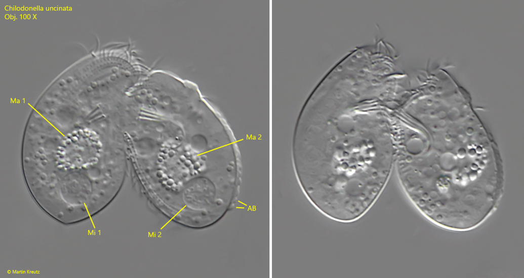
Fig. 9 a-b:Chilodonella uncinata. L = 30-33 µm. Two specimens in conjugation. Note the enlarged micronuclei (Mi 1, Mi 2) with condensed DNA filements to start the meiotic division. AB = adhering bacteria on the pellicle, Ma 1, Ma 2 = macronuclei. Obj. 100 X.