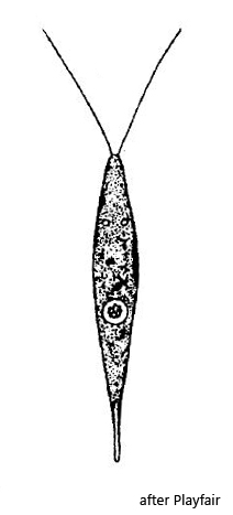
In March 2020 I found a mass development of Chlorogonium elegans in a shallow pool of water at Mindelsee. The cells were very sensitive and shed off the flagella at the slightest coverslip pressure. Therefore I could only photograph freely swimming specimens. The assignment of the Chlorogonium species is not easy, since many of the species described in the literature have turned out to be invalid. In the present case the absence of a pyrenoid was an important feature and the chloroplast not being spirally coiled. This eliminates many, potentially eligible species. The cells in my population had a body length of 38–44 µm and the flagella were slightly half the length of the spindle-shaped cell. Finally, this combination of features leads to the species Chlorogonium elegans.
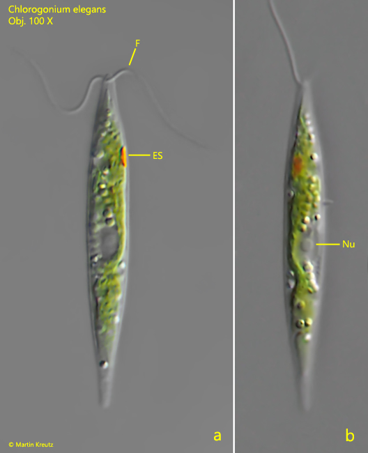
Fig. 1 a-b: Chlorogonium elegans. L = 42 µm. A freely swimming specimen in two focal planes. Note the absence of a pyrenoid. ES = eyespot, F = flagella, Nu = nucleus. Obj. 100 X.
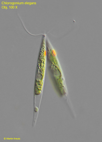
In the samples with Chlorogonium elegans there were very many specimens which were in the process of conjugation. Conjugation can be easily distinguished from asexual cell division, because the latter occurs by transverse division.
Fig. 2: Chlorogonium elegans. A fused pair of cells during conjugation. Obj. 100 X.