cells spherical, subspherical or hemispherical (after division)
cells 48–65 µm (without sheath)
two-cell colonies 80–107 µm in diameter (without sheath)
colonies of 2–4 cells, no larger aggregates
layered sheath (after each division a new layer)
sheath colorless, tightly fitting
color of cells olive green, grayish-blue or violet, never bluegreen
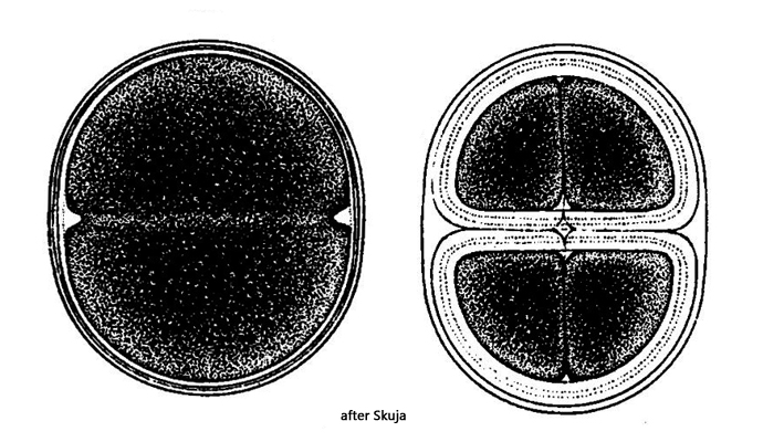
Chroococcus giganteus
Chroococcus giganteus is one of the cyanobacteria with the largest cells. Colonies are usually found with 2 or 4 cells and the division stages in between. Round single cells can only be found very rarely.
The appearance of Chroococcus giganteus is very characteristic, but it is difficult to distinguish it from the similar species Chroococcus turgidus. The only distinguishing feature seems to be the size of the individual cells and the color of the cells. The individual cells of Chroococcus turgidus are reorted to measure up to 32 µm and are clearly blue-green in color. For the cells of Chroococcus giganteus, the specification 54–65 µm is repeatedly found in the literature. This range was adopted from the first description by West (1892) and has been copied by the later authors. A redescription of the Chroococcus giganteus by Skuja (1956) is available, in which he extends the range to 48-65 µm for a single cell. Finally, John et al. (2002) postulate that Chroococcus turgidus and Chroococcus giganteus are possibly one species because transitional forms have also been found which lie between the two size ranges.
The semi-circular cells in my population were 40-52 µm wide in the two-cell colonies. They are therefore between the stated sizes of the two species. Since the cells of all specimens I found measured at least 40 µm and the color of the cells was rather olive green, but not blue-green, I stick to the identification Chroococcus giganteus.
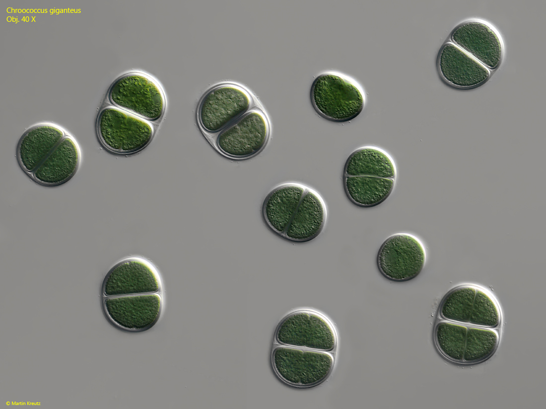
Fig. 1:Chroococcus giganteus. Overview of specimens in different cell division states. Obj. 40 X.
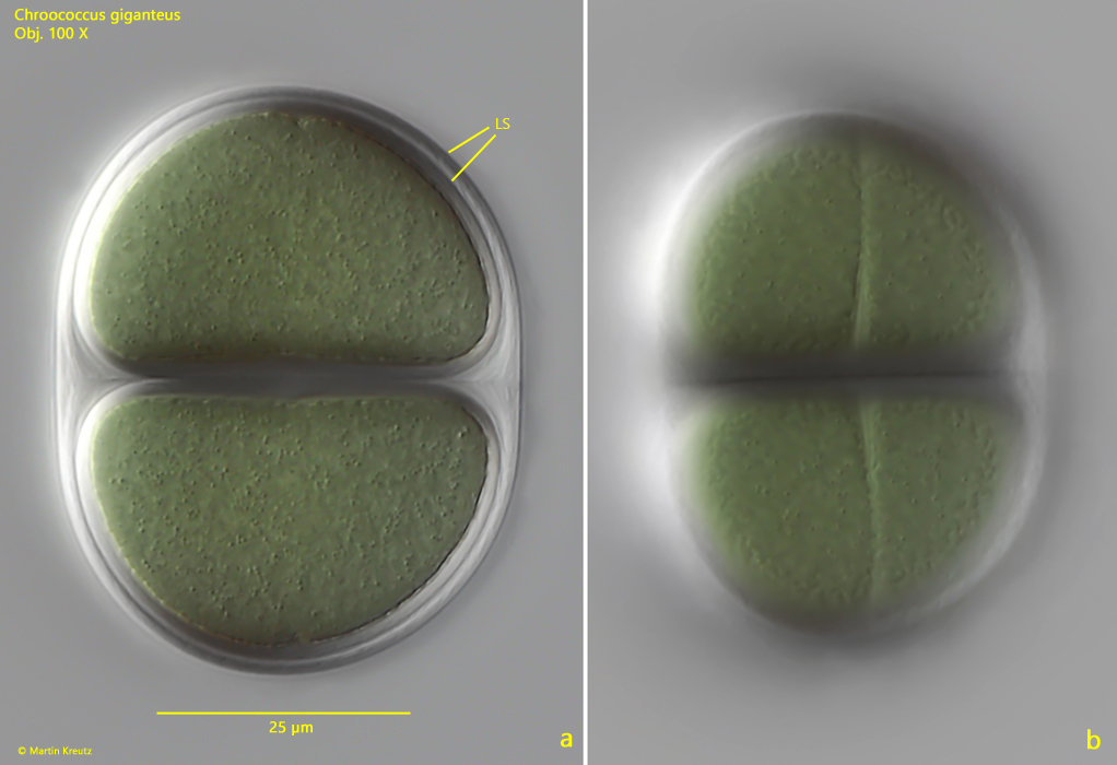
Fig. 2 a-b:Chroococcus giganteus. L = 56 µm (longitudinal axis, without sheath). An unsquashed two-cell colony during cell division (b). LS = layered sheath. Obj. 100 X.
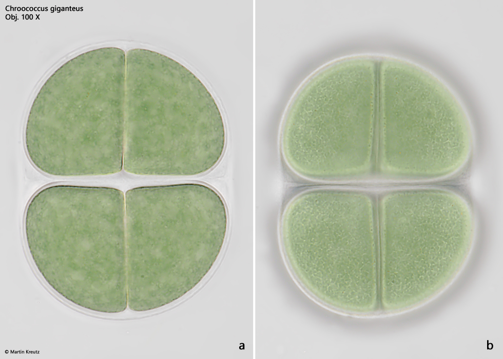
Fig. 3 a-b:Chroococcus giganteus. L = 56 µm (longitudinal axis, without sheath). A second unsquashed colony at the stage of almost finished cell division. Obj. 100 X.
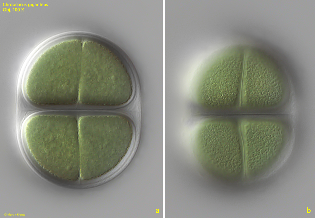
Fig. 4 a-b:Chroococcus giganteus. L = 72 µm (longitudinal axis, without sheath). A four-cell colony in brightfield illumination. Obj. 100 X.