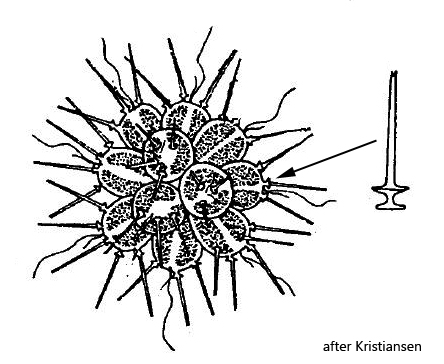
at the anterior end of each cell two cup-shaped scales with an attached long, hollow rod
Chrysosphaerella brevispina
This chrysophyte is described as common and widespread. But I could find only one specimen 2022 in the Simmelried. Possibly I have often overlooked it, because it resembles Synura at low magnifications. However the hollow protruding rods are characteristic. It requires high magnifications to reveal the complicated structure of the silicon scales. At the end of each cell two specially shaped scales are located, which can best be described as cup-shaped. The long hollow rods sit on the edges of these “cups”, but are not firmly connected to them. At low mechanical stress (cover glass pressure) the hollow rods detach.
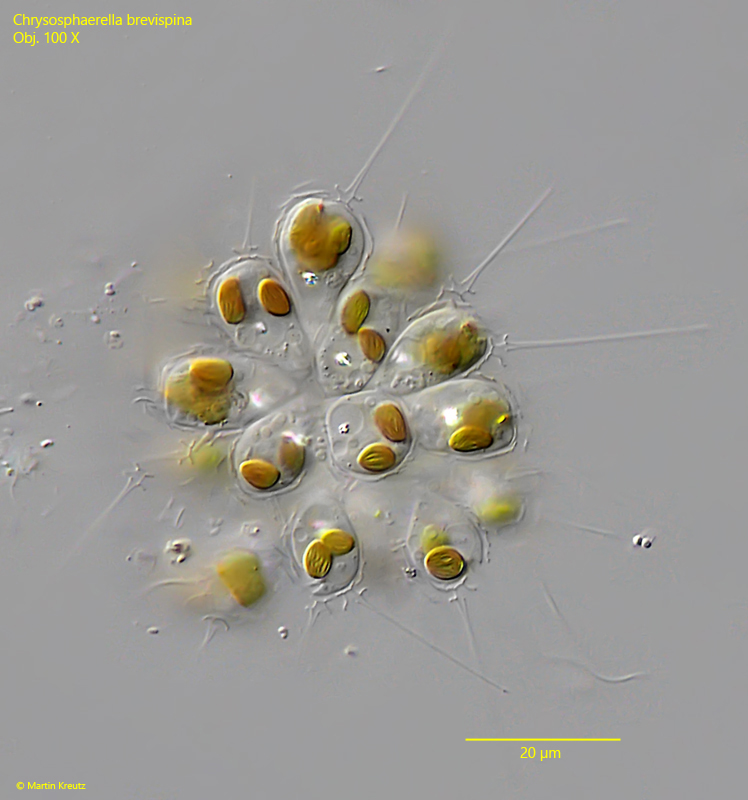
Fig. 1: Chrysosphaerella brevispina. d = 42 µm. A slightly squashed colony. Obj. 100 X.
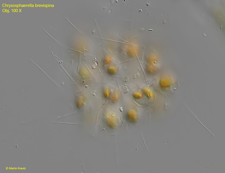
Fig. 2: Chrysosphaerella brevispina. d = 42 µm. A slightly squashed colony. Focal plane on the colony surface. Obj. 100 X.
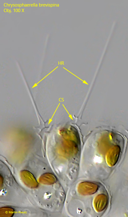
Fig. 3: Chrysosphaerella brevispina. At the anterior end of each cell two cup-shaped scales (CS) are located. To each of these scales hollow rods (HR) are attached. This delicate structure disintegrates with increasing cover glass pressure. Obj. 100 X.