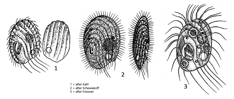
oral apparatus located at posterior end, left side
spindle-shaped extrusomes about 3 µm long
contractile vacuole sub-terminal
globular macronucleus positioned centrally with one adjacent micronucleus
about 4–5 caudal cilia
Cinetochilum margaritaceum
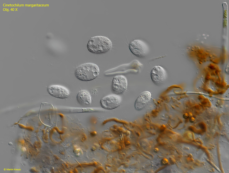
Cinetochilum margaritaceum is one of the most common ciliates besides Cyclidium, Paramecium or Coleps. Although it is so common and can occur in masses, it is not a particularly easy target for photography because it is only 15–40 µm in size, usually staying below 30 µm. Therefore, there are not as many light microscopic examinations as one would think. Under the coverslip, Cinetochilummargaritaceum is not a fast swimmer and likes to gather around particles of detritus, where the ciliate feeding on bacteria (s. fig. 1).
Fig. 1:Cinetochilum margaritaceum. Several specimens feeding on bacteria around a detritus flake. Obj. 40 X.
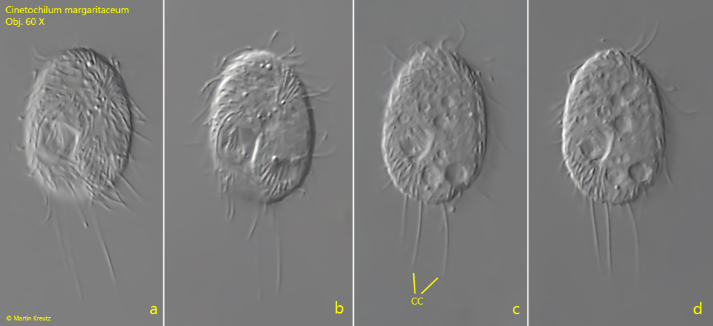
Fig. 2 a-d:Cinetochilum margaritaceum. L = 24 µm. Ventral view of a freely swimming specimen. CC = caudal cilia. Obj. 60 X.
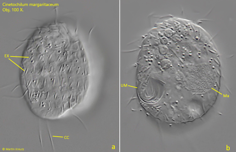
The ventral side of Cinetochilum margaritaceum is finely furrowed. The furrows originate apically and run across the cell body in counterclockwise direction. The oral apparatus is located posteriorly at the right side. The oral opening is almost rectangular in optical section. The right side of the mouth opening is encompassed by the undulating membrane (UM). The contractile vacuole (CV) is located to the left of the oral apparatus at the posterior end. In my population of Cinetochilum margaritaceum the macronucleus (Ma) was not located centrally but in the posterior third of the cell (s. fig. 3 d).
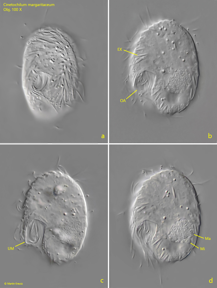
Fig. 3 a-d: Cinetochilum margaritaceum. L = 25 µm. slightly pressed specimen, ventral view, focus on oral apparatus. EX = extrusomes, Ma = macronucleus, Mi = micronucleus, OA = oral apparatus, UM = undulating membrane. Obj. 100 X.
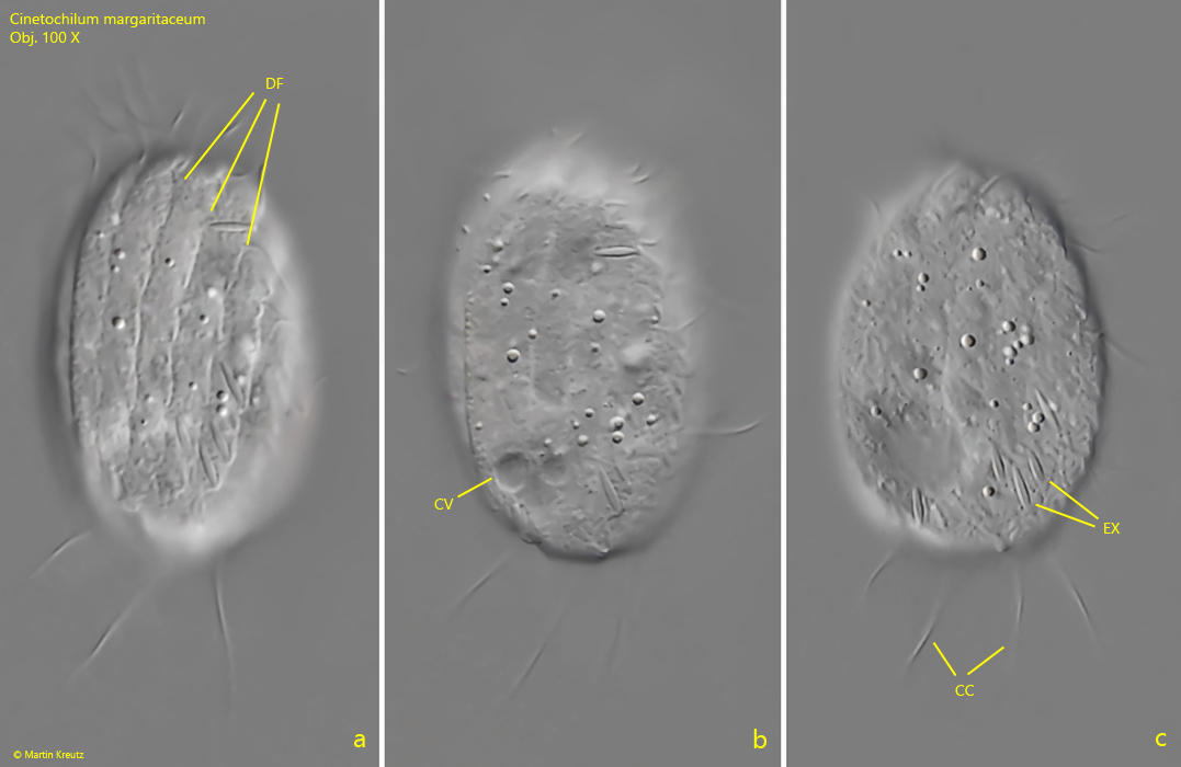
The dorsal side is wider and more deeply furrowed. The furrows run clockwise. Between the furrows 2–3 µm long extrusomes (EX) are intercalated. The furrows run to the posterior end where they form a notched posterior margin. From these notches caudal cilia (CC) arise which are about 15–20 µm long. According to my observations there are 4 in each cell. Literature sources indicate a range of 3–5 caudal cilia.
Fig. 4 a-c:Cinetochilum margaritaceum. L = 25 µm. Different focal planes of a slightly squashed specimen from dorsal. CC = caudal cilia, CV = contractile vacuole, DF = dorsal furrows, EX = extrusomes. Obj. 100 X.
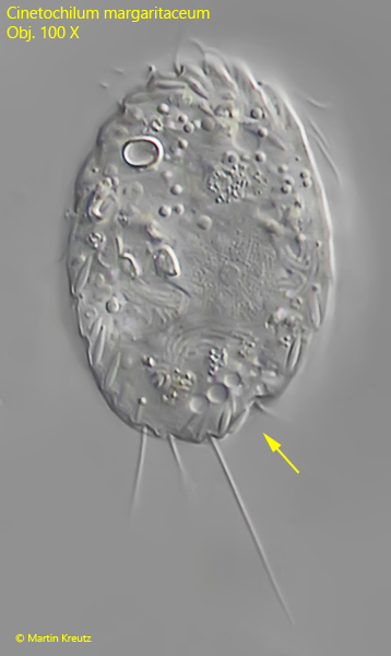
Fig. 5:Cinetochilum margaritaceum. L = 35 µm. Ventral view of a slightly squashed specimen. Note the oval shape of the cell with the characteristic indentation at the posterior right margin (arrow). Obj. 100 X.
Fig. 6 a-b:Cinetochilum margaritaceum. L = 29 µm. Dorsal view (a) and ventral view (b) of a slightly squashed specimen. Note the rows of extrusomes (EX) arranged between the dorsal furrows. Ma = macronucleus, UM = undulating membrane. Obj. 100 X.
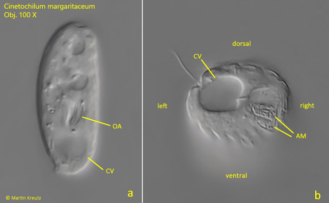
The oval body of Cinetochilum margaritaceum is distinctly dorso-laterally flattened, which can be very well seen in lateral view (s. fig. 7 a). In a view from posterior the adoral membranelles in the apparatus are visible (s. fig. 7 b).
Fig. 7 a-b:Cinetochilum margaritaceum. Lateral view (a) and view from posterior (b) of a freely swimming specimen. AM = adoral membranelles, CV = contractile vacuole, OA = oral apparatus. Obj. 100 X.
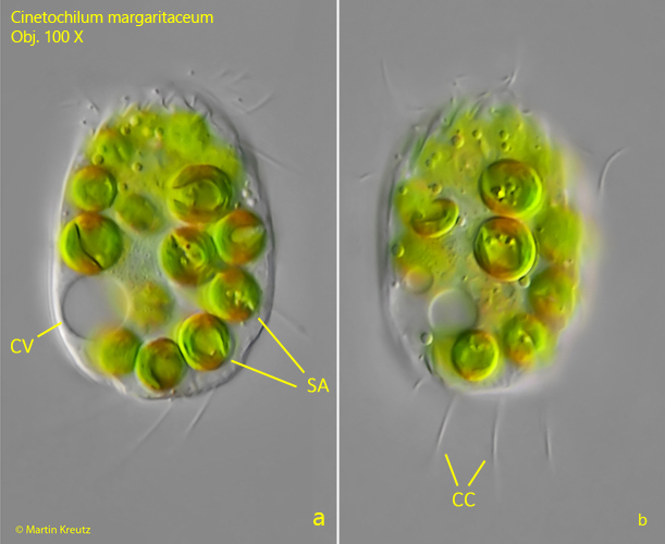
Kahl also describes a form of Cinetochilum margaritaceum with symbiotic algae. I found this form rarely but regularly in Simmelried as well as in the Purren pond. The green form of Cinetochilum margaritaceum contains 15–20 symbiotic algae (Ø = 6 µm) and is somewhat larger with a distinctly different behavior compared to the colorless form. While the colorless form moves in small circles, the green form swims around quite fast and hardly ever rests. In addition it is sensitive to pressure of the coverslip. Considering the differences to the colorless form (size, symbiotic algae and behavior) Cinetochilum margaritaceum with symbiotic algae can, in my opinion, be defined as a separate species.
Fig. 8 a-b:Cinetochilum margaritaceum. L = 34 µm. Ventral view of a specimen with symbiotic algae (SA). CC = caudal cilia, EX = extrusomes, OA = oral apparatus. Obj. 100 X.
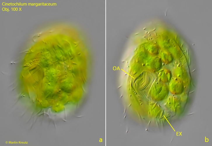
Fig. 9 a-b:Cinetochilum margaritaceum. L = 34 µm. A specimen with symbiotic algae from ventral with focal plane on the oral apparatus (OA). EX = extrusomes. Obj. 100 X.
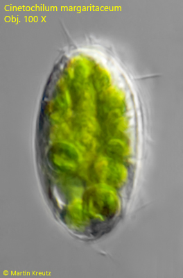
Fig. 10:Cinetochilum margaritaceum. L = 34 µm. A freely swimming specimen with symbiotic algae from lateral. Obj. 100 X.