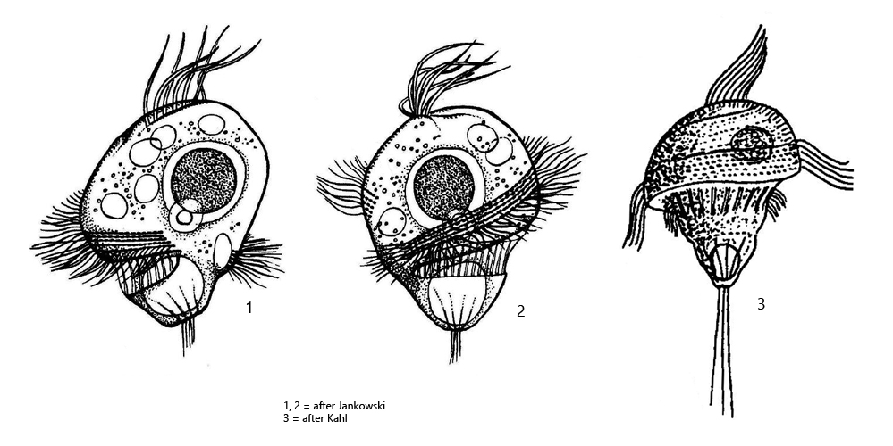
perizonal stripe begins dorsal and runs spirally around body
adoral zone parallel to perizonal stripe
somatic ciliation absent
mouth opening in posterior third
globular macronucleus with adjacent micronucleus
contractile vacuole terminal
tuft of caudal cilia with body length
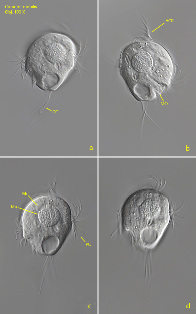
Cirranter mobilis
Cirranter mobilis was first described by Penard (1922) as Trochella mobilis. This name was adopted by Kahl. Jankowski then transferred the species to the genus Cirranter in 1964. The genus Cirranter belongs to the Apometopidae (Foissner, 2016). The perizonal stripe of Apometopidae consists of only 4 rows of cilia.
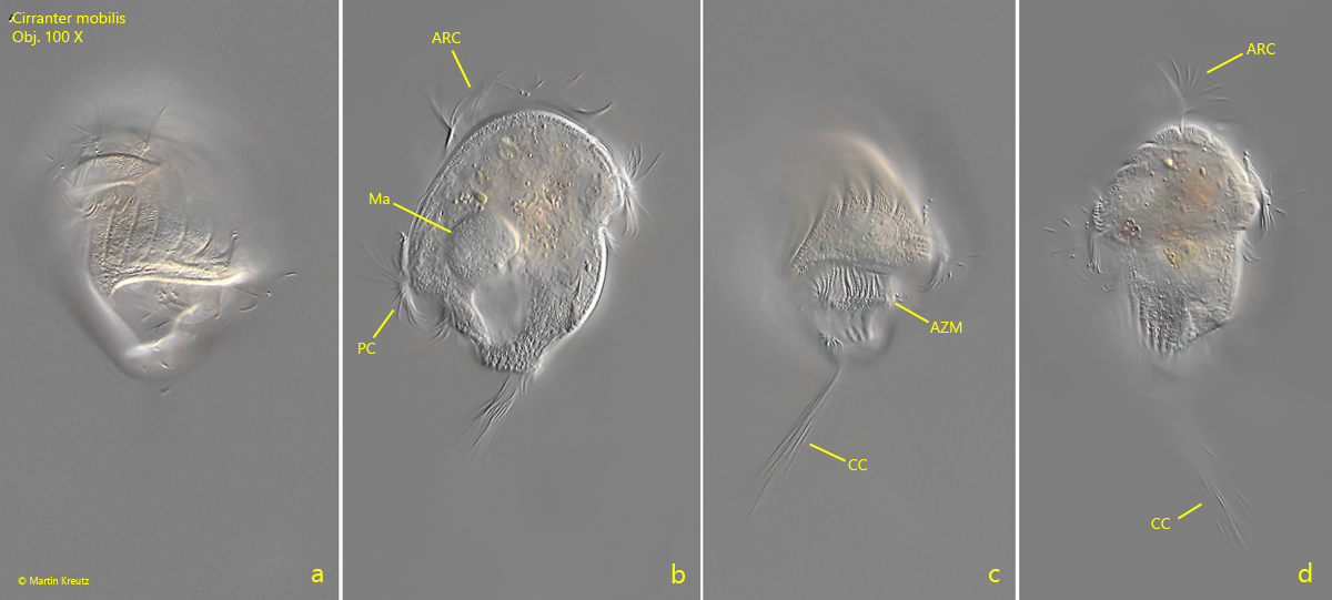
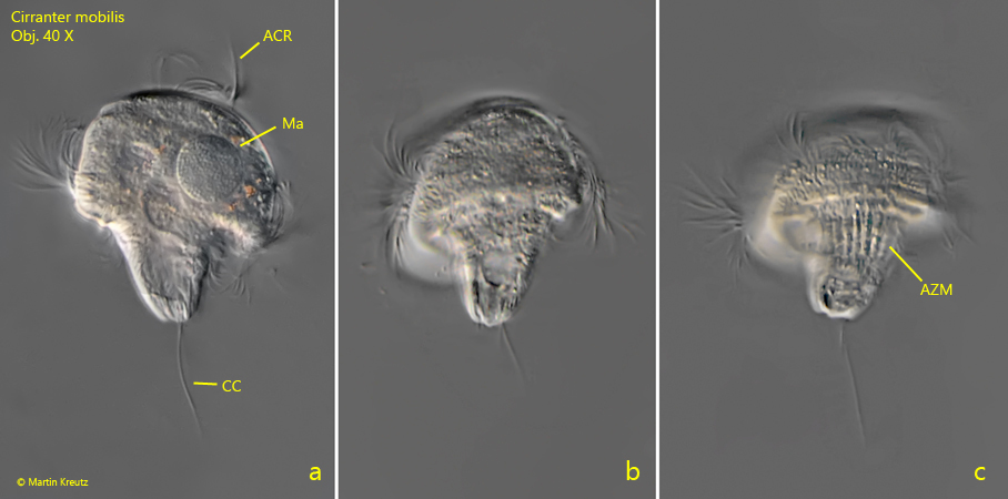
So far I have found Cirranter mobilis exclusively in Simmelried where I find the species regularly and throughout the year. Although the ciliate in my population usually grows only 30 – 35 µm, I recognize it by the apical row of cilia (s. figs. 1 b, 2 b, 2 d, 3 a and 4 b) and by the tufts of long caudal cilia (s. figs. 2 c and 2 d). At higher magnification, one can also see the ridges between the membranelles of the adoral zone (s. fig. 3 c), which is also a characteristic feature of this species. Cirranter mobilis is a fast swimmer (hence the name) and is not easy to photograph because it is also very coverslip sensitive, which Kahl already noted. Kahl helped himself by leaving the slide with the sample in a humidity chamber for a day. By this treatment the specimens should become slower. I did not try this approach because the microflash also freezes fast moving objects. However, this method was not available to Kahl.
Fig. 1 a-d:Cirranter mobilis. L = 31 µm. A freely swimming specimen from left (a, c, d) and right (b). Note the apical row of cilia (ACR). CC = caudal cilia, Ma = macronucleus, Mi = micronucleus, MO =mouth openung, PC = perizonal cilia. Obj. 100 X.
Fig. 2 a-d:Cirranter mobilis. L = 30 µm. A second freely swimming specimen from dorsal (a), ventral (b, c) and from right (d). ACR = apical row of cilia, AZM = adoral zone of membranelles, CC = tuft of caudal cilia, Ma = macronucleus, Mi = micronucleus. Obj. 100 X.
Fig. 3 a-c:Cirranter mobilis. L = 47 µm. A third freely swimming specimen from dorsal (a) and ventral (b, c). Note the “ribs” between the membranelles of the adoral zone (AZM). ACR = apical row of cilia, CC = caudal cilia, Ma = macronucleus. Obj. 100 X.
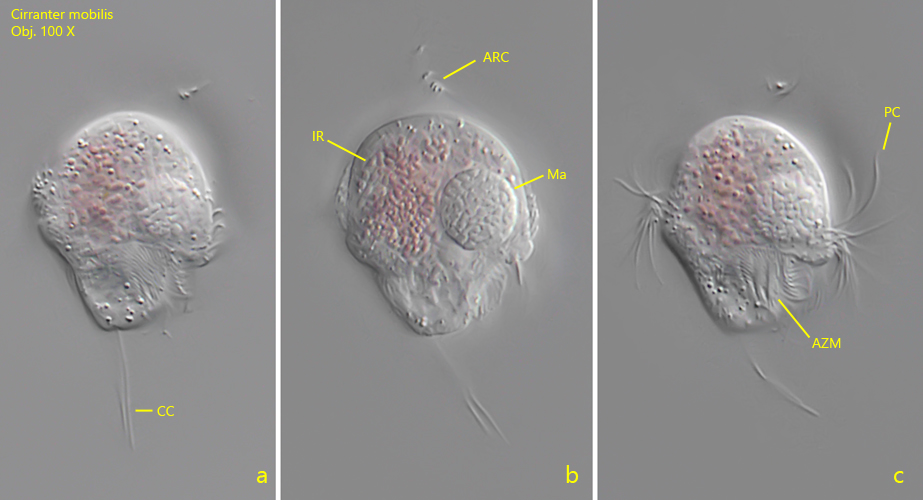
Fig. 4 a-c:Cirranter mobilis. L = 28 µm. A freely swimming specimen with ingested rhodobacteria (IR) from ventral. ACR = apical row of cilia, AZM = adoral zone of membranelles, CC = tuft of caudal cilia, Ma = macronucleus, PC = perizonal cilia. Obj. 100 X.
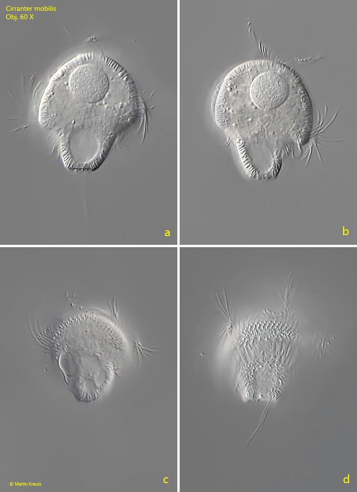
Fig. 5 a-d:Cirranter mobilis. L = 40 µm. Different views of a freely swimming specimen. Obj. 60 X.
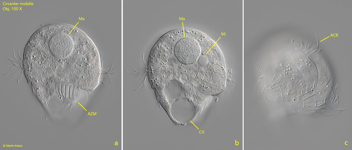
Fig. 6 a-c:Cirranter mobilis. L = 45 µm. A freely swimming specimen from ventral (a, b) and from apical (c). ACR = apical row of cilia, CV = contractile vacuole, Ma = macronucleus, Mi = micronucleus. Obj. 100 X.