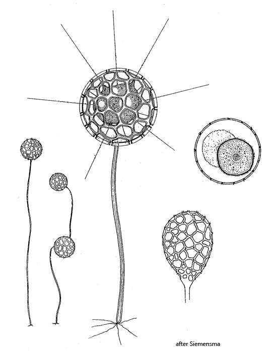
body spherical in an organic test with polygonal pores
granulated axopodia protruding through the pores
test spherical or oval shaped, usually on a long, hollow stalk
length of test 30–90 µm
protoplast does not fill entire test
solitary or in colonies
nucleus central with large nucleolus
one or more contractile vacuoles
Clathrulina elegans
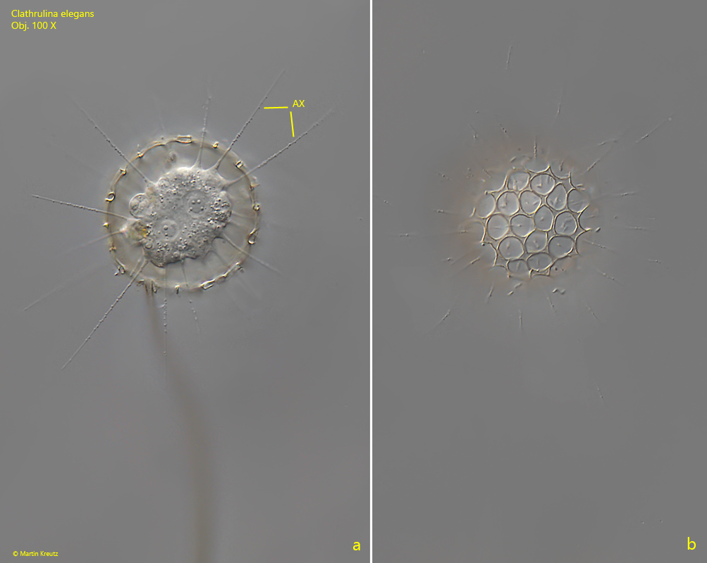
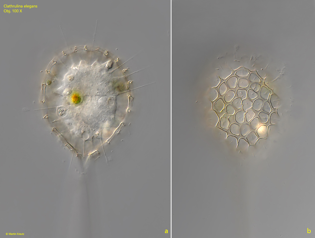
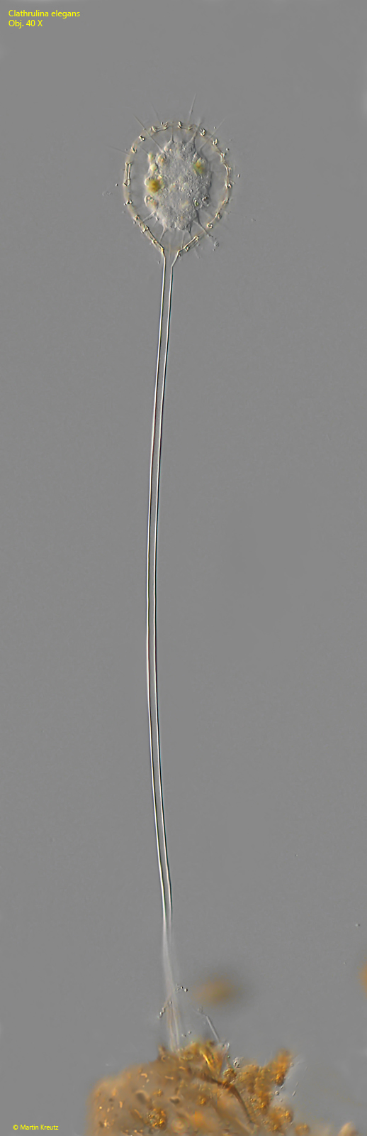
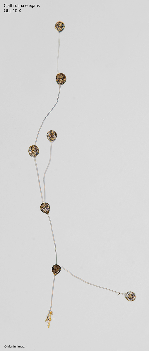
I find Clathrulina elegans regularly, but not very often. The characteristic test with polygonal pores makes it easy to identify the species. I find mostly solitary specimens on a stalk (s. fig. 3). The length of the stalk is very variable. However, the stalk is always hollow. The shape of the test is also variable. It can be spherical (s. fig. 1 a-b) or oval (s. fig. 2 a-b). The pores in the test are approximately polygonal with rounded corners. Under coverslip pressure the test breaks. It is therefore probably not purely organic. The axopodia, which protrude through the pores, are granulated and not very long (about 10–40 µm). Only rarely have I found colonies (s. fig. 4). These are formed when the biflagellated swarmers leave the test through the pores and attach themselves directly to the test of the mother cell to form a new stalk and test. When this process is repeated often, apparently branched colonies are formed.
Fig. 1 a-b:Clathrulina elegans. D = 48 µm (of test). Two focal planes of a specimen in a spherical test. In the protoplast two nuclei are visible. AX = axopodia. Obj. 100 X.
Fig. 2:Clathrulina elegans. L = 65 µm (of test). A second specimen in an oval shaped test. Obj. 100 X.
Fig. 3:Clathrulina elegans. L = 465 µm (of test and stalk). A solitary specimen on a 400 µm long, hollow stalk. Obj. 40 X.
Fig. 4:Clathrulina elegans. L = 1500 µm (of colony). A colony of 7 specimens. Each specimen is attached with the stalk to test of an other specimen. In the tests spherical cysts are visible. Obj. 10 X.