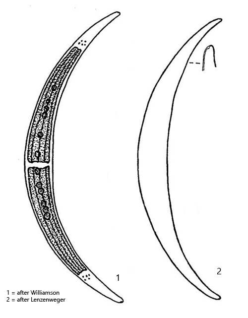
inner margin sometimes straight or slightly convex in mid-region
length 180–300 µm, width 20–30
cell wall smooth without striation
two chloroplasts, each with 3–5 longitudinal ridges
several pyrenoids arranged along cell axis
girdle bands absent, sometimes pseudo-girdle bands
apices with each one vacuole filled with crystals
nucleus central
Closterium dianae
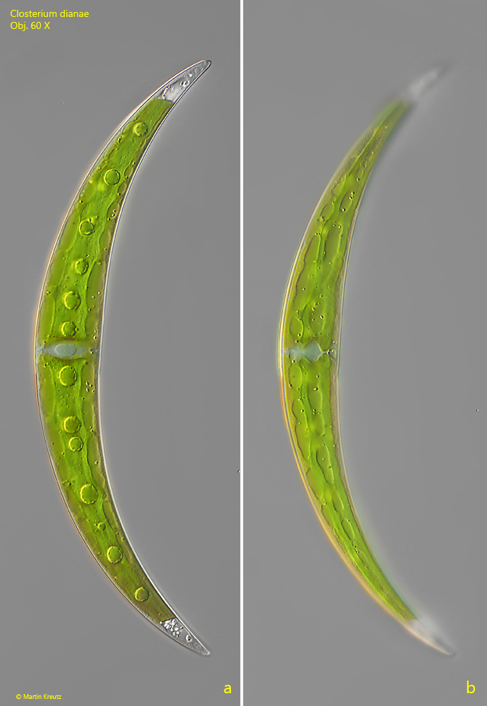
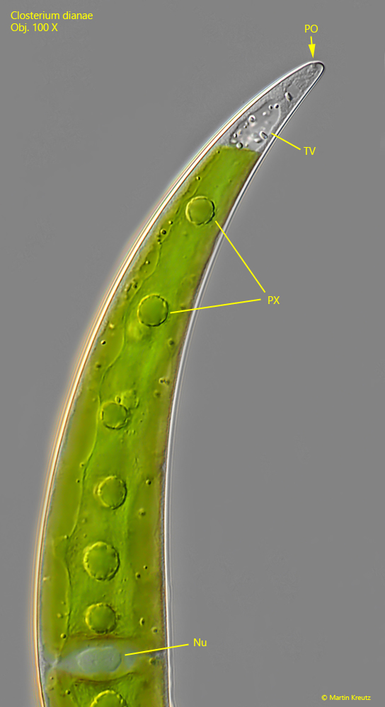
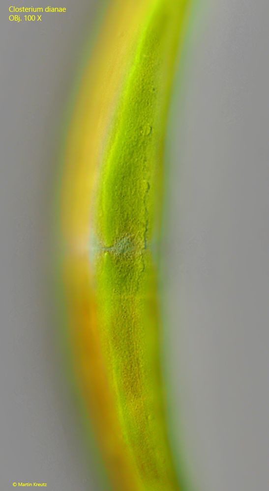
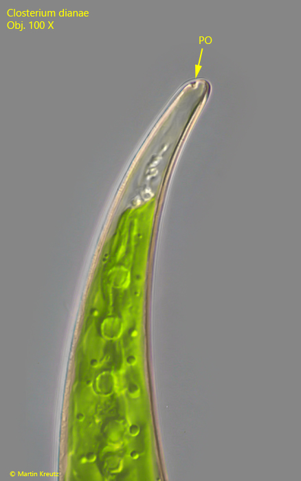
I have only rarely found Closterium dianae so far. The cells are slender and quite evenly curved. Essential features for identification are the pores at the apices (s. fig. 2), the smooth cell wall without striations (s. fig. 3) and the pyrenoids arranged along the longitudinal axis (s. figs. 1 a and 2).
Fig. 1 a-b:Closterium dianae. L = 220 µm. Two focal planes of a young specimen with a transparent cell wall. Obj. 60 X.
Fig. 2:Closterium dianae. Focal plane on one of the apices with the terminal vacuole (TV) filled with bariumsulfate crystals and the porus (PO). Nu = nucleus, PY = pyrenoids. Obj. 100 X.
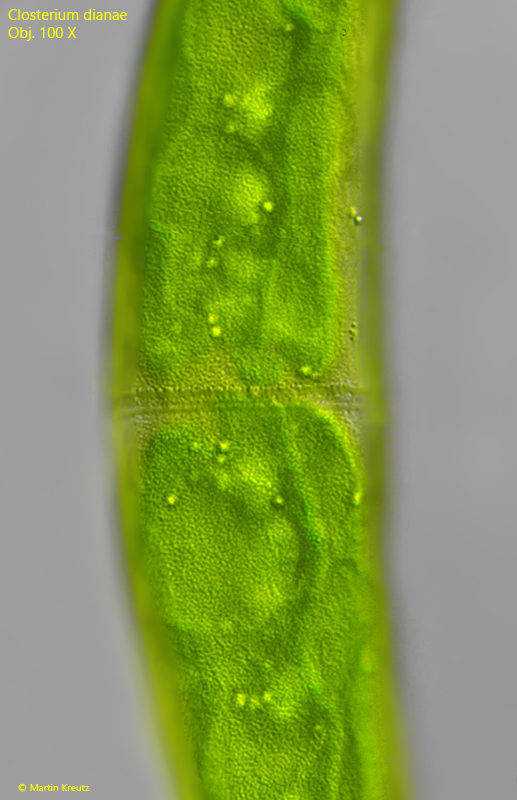
Fig. 3:Closterium dianae. The cell wall is smooth without a striation. Obj. 100 X.
Fig. 4:Closterium dianae. The porus (PO) in the apex of a second specimen. Obj. 100 X.
Fig. 5:Closterium dianae. The granulated cell wall of a second specimen. Obj. 100 X.