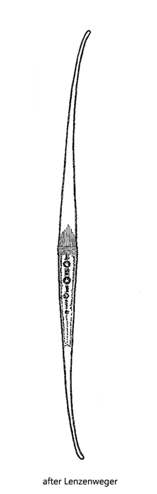
cell body attenuating into long narrow ends, mid-body spindle-shaped
length 270–550 µm, width 18–30 µm
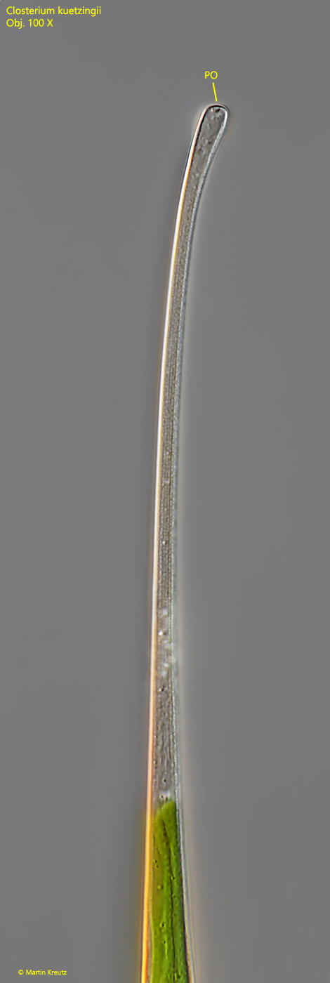
distal ends curved, slightly swollen, each with an apical porus
two chloroplasts with 5 lamellae each in both semi-cells
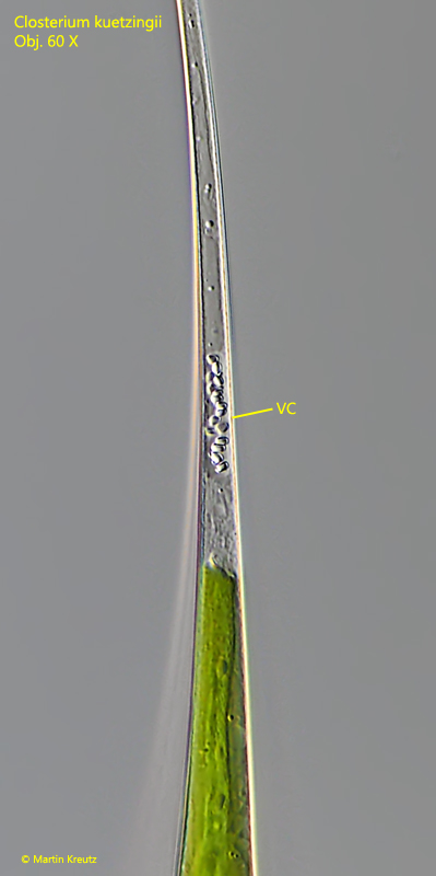
at the distal ends of the chloroplasts each one vacuole filled with oval shaped crystals
each chloroplast with 4–6 pyrenoids
cell wall brownish, with striation of 8–11 lines/10 µm
nucleus central between the chloroplasts
Closterium kuetzingii
I find Closterium kuetzingii regularly in the Simmelried. The shape of the cell is somewhat reminiscent of an arch. The ends of the cells are slightly bent. Closterium kuetzingii can be distinguished from the similar species Closterium rostratum and Closterium setaceum by the shape and length of the semi-cells. In Closterium setaceum, the distal ends are much more strongly elongated and slender, while Closterium rostratum is more compact in appearance and has shorter and thicker distal ends.
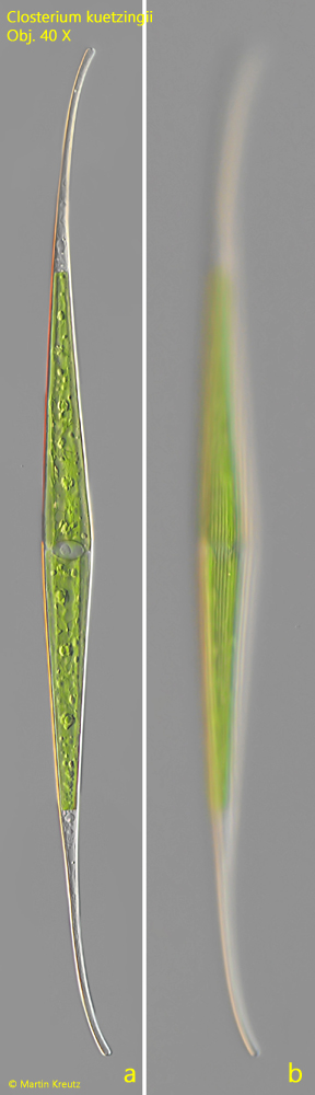
Fig. 1 a-b:Closterium kuetzingii. L = 465 µm. Two focal planes of a slightly squashed specimen. Obj. 40 X.
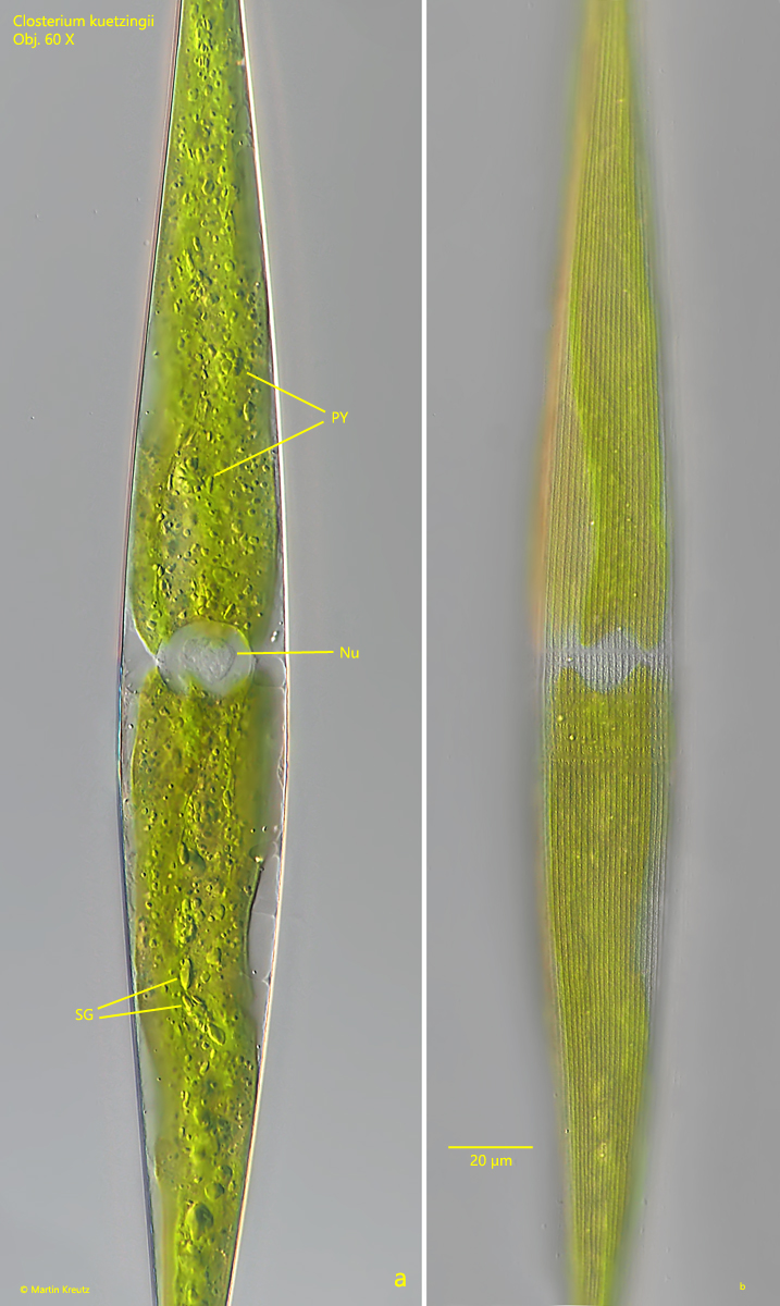
Fig. 2 a-b:Closterium kuetzingii. L = 578 µm. Two focal planes of the spindle-shaped mid-body. Nu = nucleus, PY = pyrenoids, SG = starch grains. Obj. 60 X.
Fig. 3:Closterium kuetzingii. At the distal end of each chloroplast a vacuole filled with oval crystals (VC) is located. Obj. 60 X.
Fig. 4:Closterium kuetzingii. The distal end of a cell in detail. Note the slightly swollen distal end with an apical porus (PO). Obj. 100 X.