two chloroplasts, each with 3–6 longitudinal ridges
2–10 pyrenoids arranged along cell axis, sometimes scattered
girdle bands absent
apices with each one vacuole filled with oval crystals
cell wall with fine striation (14–20 striae/10 µm)
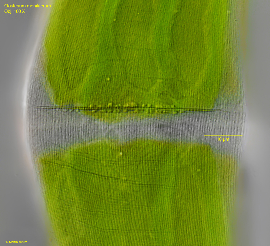
nucleus central in a cytoplasm bridge between the chloroplasts
Closterium moniliferum
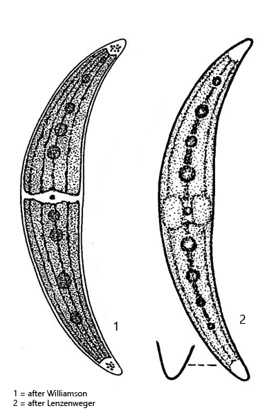
Closterium moniliforme is one of the most common representatives of the genus Closterium. I find this species in almost all my sampling sites. Closterium moniliferum can be recognized by the slightly convex shape of the ventral side and that the pyrenoids are arranged along the longitudinal axis of the half-cells. Sometimes, however, the pyrenoids can also be slightly irregularly offset to the longitudinal axis, as in the specimen shown below (s. figs. 1 a and 3). The cell wall is finely striated (s. fig. 4) and the apices are rounded and only slightly bent dorsally (s. fig. 2 a-b). In the terminal vacuoles there are numerous oval crystals of barium sulphate with unknown function (s. fig. 2 b). The similar species Closterium ehrenbergii is larger (> 400 µm) and the pyrenoids in this species are scattered on the surface of both chloroplasts.
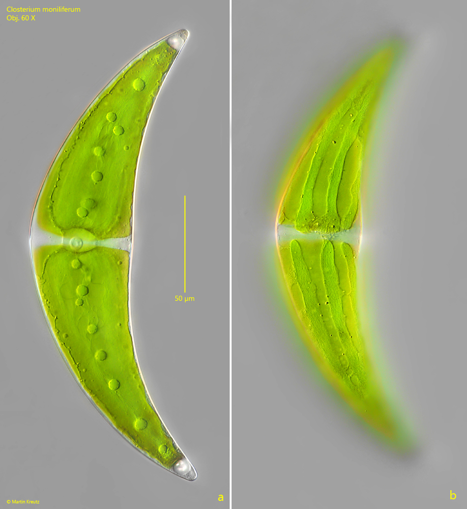
Fig. 1 a-b:Closterium moniliferum. L = 240 µm. Focal plane on the pyrenoids arranged along the cell axis (a) and on the longitudinal ridges of the chloroplasts in the half-cells (b). Obj. 60 X.
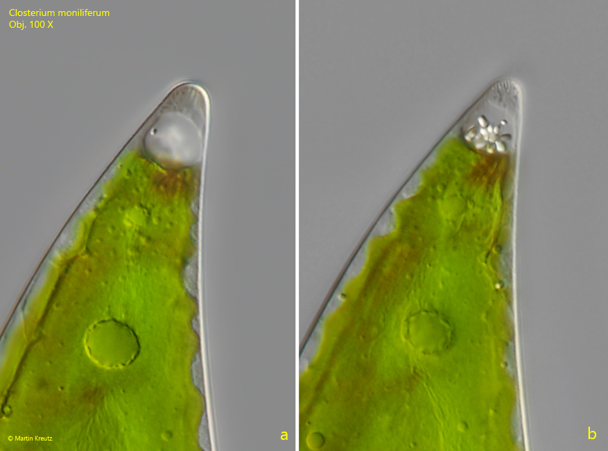
Fig. 2 a-b:Closterium moniliferum. Two focal planes on one of the apices with the terminal vacuole filled with oval bariumsulfate crystals. Obj. 100 X.
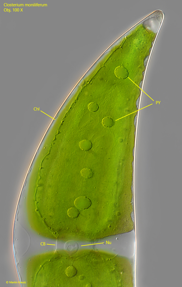
Fig. 3:Closterium moniliferum. A half-cell in detail. Note the nucleus (Nu) located between the two chloroplasts (Chl) in a cytoplasm bridge (Cb). PY = pyrenoids. Obj. 100 X.
Fig. 4:Closterium moniliferum. Focal plane on the fine striation of the cell wall. This specimen has 17 striae/10 µm. Obj. 100 X.