cell wall often brownish with 4–10 ridges per 10 µm
two chloroplasts, each with 6–9 longitudinal ridges
4–15 pyrenoids per chloroplast
girdle bands present
apices with each one vacuole filled with a single aggregate of crystals or several crystals
nucleus central
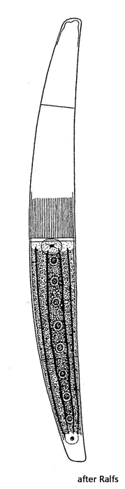
Closterium striolatum
I rarely find Closterium striolatum in the Simmelried. A sample from a bog pond near the Warnsdorfer spring (Tharandter Wald, Dresden) contained masses of the species in April 2024.
Closterium striolatum is usually longer than 300 µm and therefore stands out in the samples. The cell wall is often strongly brown in color with iron and manganese deposits. The shape of the chloroplast is striking, with longitudinal ribs (stellate type). These ribs can be connected to each other like a net (s. figs. 4 and 5). In the terminal vacuoles I found clusters of crystals partly arranged in layers (s. fig. 8 a-b).
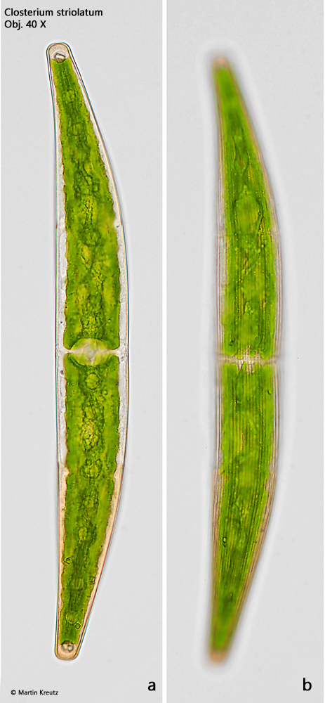
Fig. 1 a-c:Closterium striolatum. L = 328 µm. Three focal planes of a slightly squashed specimen. Obj. 40 X.
Fig. 2 a-b:Closterium striolatum. L = 328 µm. The same specimen as shown in fig. 1 a-c in brightfield illumination. Obj. 40 X.
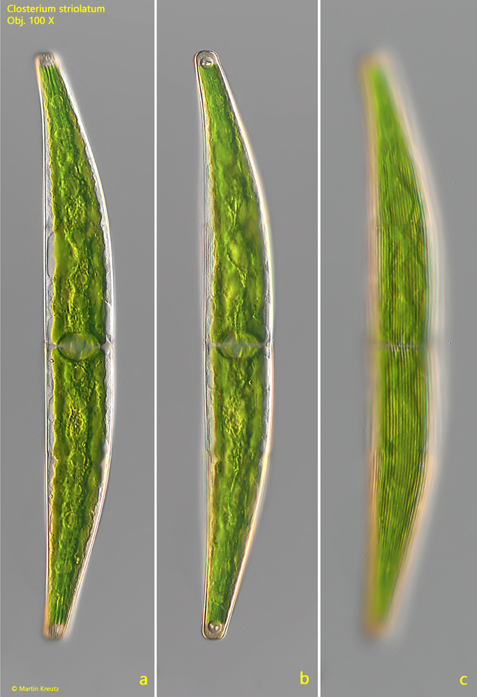
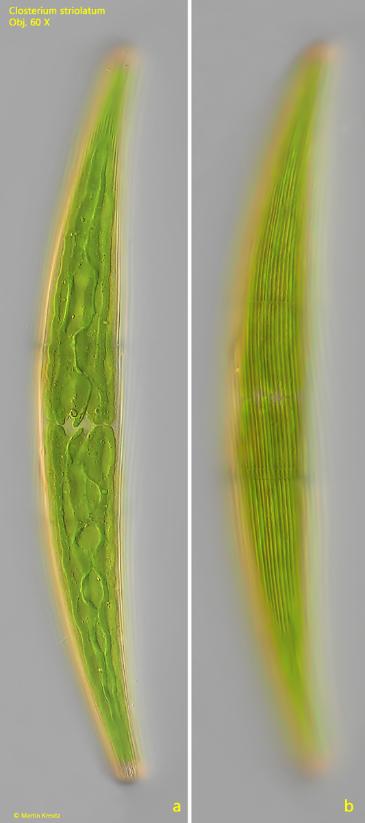
Fig. 3 a-b:Closterium striolatum. L = 305 µm. Focal plane on the ridges of the chloroplasts (a) and the striation of the cell wall (b) of a second specimen. Obj. 60 X.
Fig. 4:Closterium striolatum. The wavy longitudinal ridges of the chloroplasts. Obj. 100 X.
Fig. 5:Closterium striolatum. The longitudinal ridges of the chloroplasts (Chl) in detail with small crystals (CR) scattered between. Obj. 100 X.
Fig. 6:Closterium striolatum. The delicate striation of the punctate cell wall in detail. Obj. 100 X.
Fig. 7:Closterium striolatum. Details of the cell wall of a second specimen with suture bands (SB) and girdle bands (GB). Obj. 100 X.
Fig. 8 a-b:Closterium striolatum. The terminal vacuole (TV) with a cluster of crystals in DIC (a) and in brightfield illumination. Obj. 100 X.