one single flagellum, about three times cell length
base of flagellum surounded by circular collar of microvilli
one nucleus in anterior third with spherical nucleolus
one contractile vacuole below cell equator
Codosiga umbellata
I regularly find the branched colonies of Codosiga umbellata in several of my sampling sites, but only comparatively rarely. The colonies usually settle on dead plant material or detritus flakes. Colonies rarely settle on the floating coverslip.
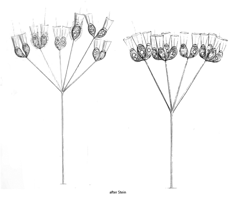
Codosiga umbellata can also be found in older literature under the synonymous names Codonocladium umbellatum and Codonosiga umbellata. Codosiga umbellata differs from the comparable species Codosiga botrys in its branched stalk. In Codosiga botrys the stalk is unbranched.
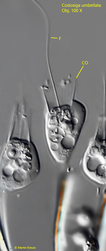
The cells at the end of the branched stalk have a rather long flagellum, which is surrounded by a ring-shaped collar at the base. (s. figs. 2 and 3). This collar is about 5–8 µm high and consists of fine filaments (microvilli, s. fig. 3). The bacteria flushed in by the movement of the flagellum remain attached to the collar and are finally phagocytized. This is why the collar is often covered with bacteria. The cells also appear to be embedded in a delicate mucus sheath, which can only be recognized in the DIC by the adhering bacteria (s. fig. 5). The cells do not form a lorica. The nucleus is located in the anterior third of the cell and has a conspicuous, spherical nucleolus. The contractile vacuole is located just below the cell equator (s. fig. 2).
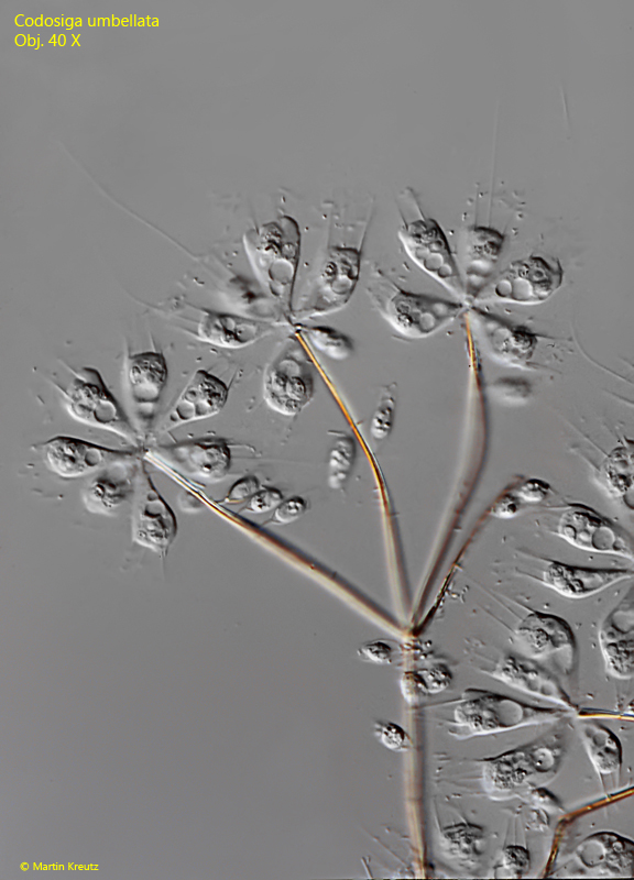
Fig. 1:Codosiga umbellata. L = 11–14 µm (of cells). A colony of about 20 cells on a brownish colored, branched stalk. Obj. 40 X.
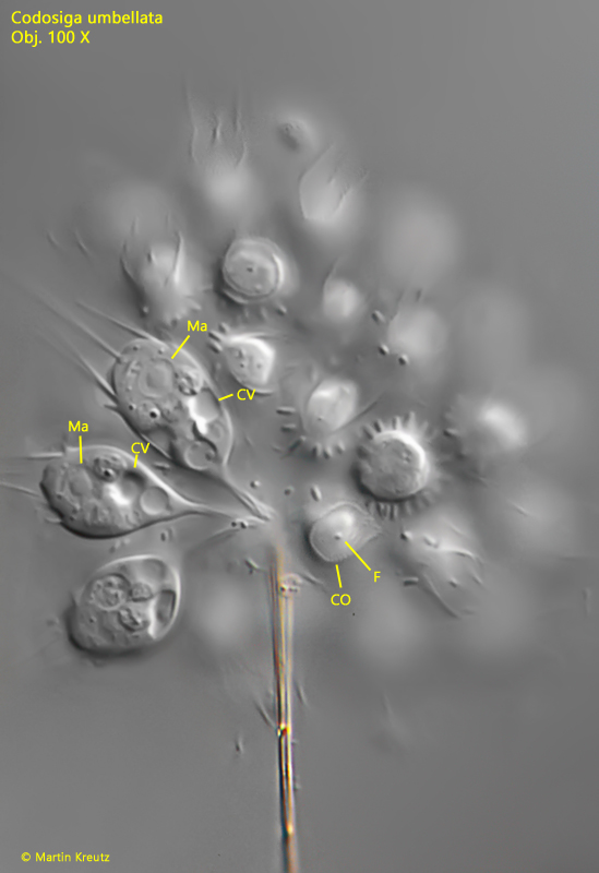
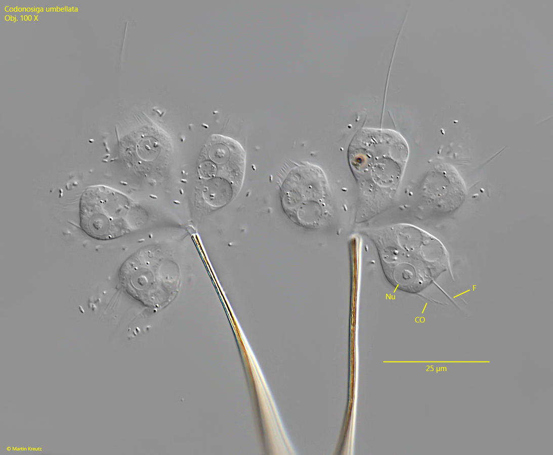
Fig. 2:Codosiga umbellata. L = 12–14 µm (of cells). A detail of a group of cells at the end of the branched stalk. Note the spherical nucleolus in the macronucleus (Ma) of the cells. CO = collar, CV = contracile vacuole, F = flagellum. Obj. 100 X.
Fig. 3:Codosiga umbellata. L = 13.8 µm. A cell in detail with strongly raised contrast for visualization of the collar (CO) composed of microvilli. F = flagellum. Obj. 100 X.
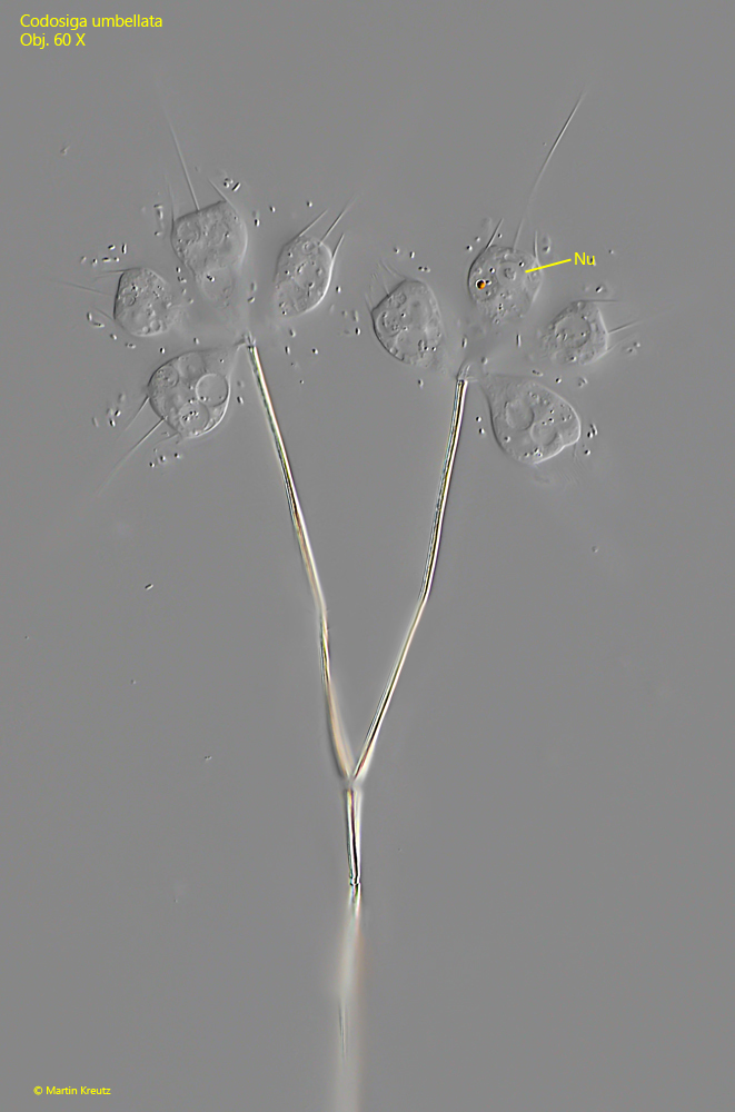
Fig. 4:Codosiga umbellata. Part of a slightly squashed colony. Nu = nucleus. Obj. 60 X.
Fig. 5:Codosiga umbellata. The colony as shown in fig. 4 in detail. The cells are covered in a delicate mucus sheath, visible by the adhering bacteria. CO = collar, F = flagellum, Nu = nucleus. Obj. 60 X.