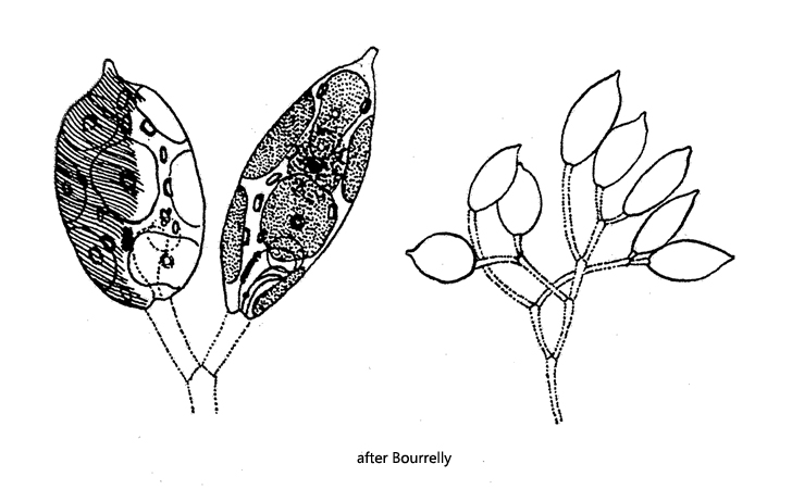
cells obvoid, rounded at anterior end, papilla at posterior end
length 28–25 µm, width 12–16 µm
6–8 disc-shaped chloroplasts, irregularly shaped
each chloroplast with a small pyrenoid
eyespot near anterior end
nucleus central
sessile cells slightly metobolic
sessile cells on dichotomously branched, mucilaginous stalks
width of hollow stalks 2–2.5 µm
motile cells with one flagellum
cells faintly striated
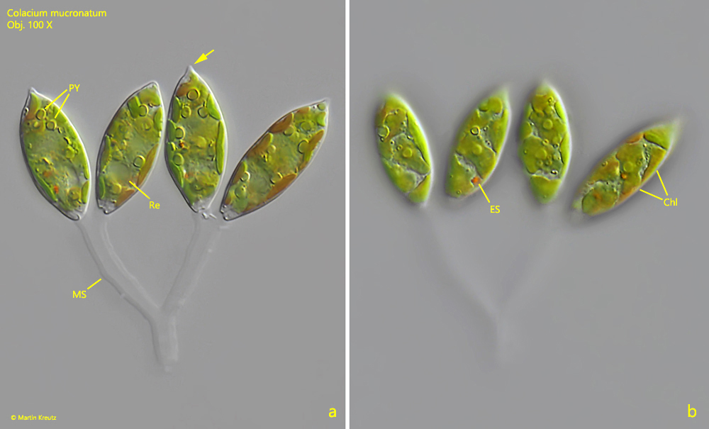
Colacium mucronatum
Colacium mucronatum belongs to the Euglenophyceae and has a sessile and motile phase in the life cycle. In the sessile phase, the cells sit with their anterior end (i.e. where the flagellum originates) on gelatinous stalks. The flagellum is fused with the cell body in this phase. On the stalks the cells divide and form branched colonies. The species Colacium mucronatum can be recognized by the dichotomous branching of the stalks as well as by the typical papilla at the posterior end. They are usually oval or slender in shape. The similar species Colacium vesiculosum also has a papilla at the posterior end, but the cell as a whole tapers to the posterior end in a cone shape. This is not the case with the species presented here.
Fig. 1 a-b:Colacium mucronatum. L = 25 – 28 µm. Two focal planes of a small colony of 4 cells. Note the characteristic papilla at the posterior end of the cell (arrow). Chl = chloroplasts, ES = eyespot, MS = mucilaginous stalk, PY = pyrenoids, Re = reservoir. Obj. 100 X.