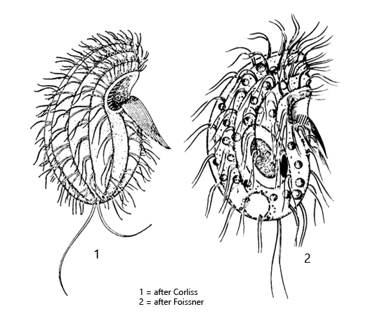
body reniform, dorsal side convex, ventral side almost straight
length 10–60 µm, usually 20–40 µm
semicircularly indented at the mouth opening
apical keel with 5–7 ribs
macronucleus ellipsoid with one central nucleolus
mouth opening with a right and left field of polykineties
cilia of the left polykinetid are long, forming a “beard”
10–13 ciliary rows of paired cilia
contractile vacuole terminal
two caudal cilia (hard to see)
Colpoda steinii
I find Colpoda steinii in almost all moss samples that I soak with rainwater in Petri dishes. After a few days, populations of varying sizes occur. However, Colpoda steinii can also be found in hay infusions, soil samples and even in plankton.
Colpoda steinii can easily be confused with other Colpoda species, such as Colpoda ecaudata or Colpoda aspera. Colpoda ecauda, however, has several nucleoli in the macronucleus and not just a central one. Colpoda aspera is smaller and has a pointed apical end. Both species also have no caudal cilia, but Colpoda steinii has.
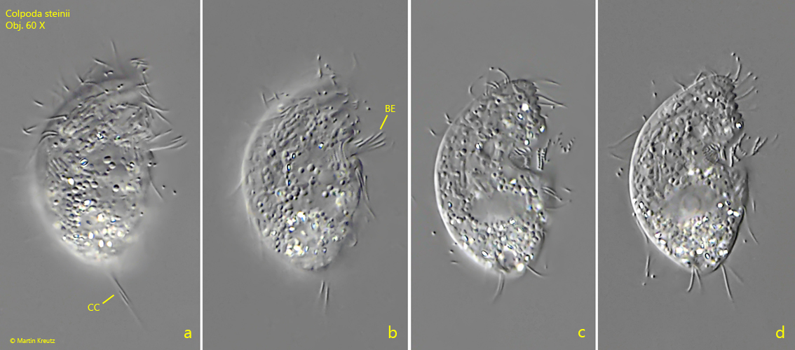
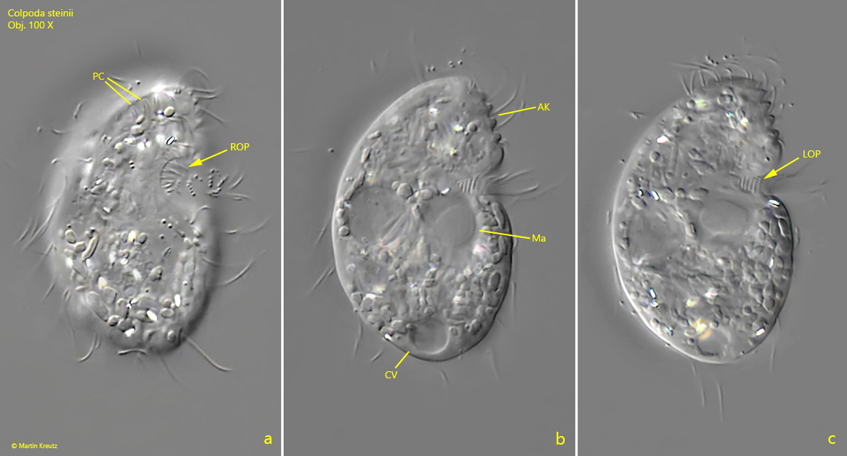
The two caudal cilia of Colpoda steinii are hard to see at high magnifications because they are constantly moving and are rarely in focus. It is therefore advisable to examine specimens at first at lower magnifications. The characteristic “beard”, which originates in the middle of the ventral side, is also striking (s. fig. 2 b). These are the long cilia of the left ciliary field of the mouth opening. The funnel-shaped mouth opening has a right and a left oral ciliary field, called right and left polykinetids. These dense fields of cilia are difficult to recognize under the light microscope. However, it is possible to distinguish between the left and right polykinetid when focusing carefully through the mouth opening of the specimen. In fig. 3 c and 4 the left oral polykinetid is visible.
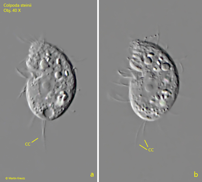
Fig. 1 a-b:Colpoda steinii. L = 28 µm. A freely swimming specimen from left. Note the two caudal cilia (CC). Obj. 40 X.
Fig. 2 a-d:Colpoda steinii. L = 33 µm. A second freely swimming specimen from right. Note the “beard” (BE) of long cilia of the left oral polykinetid. CC = caudal cilia. Obj. 60 X.
Fig. 3 a-c:Colpoda steinii. L = 39 µm. A slightly squashed specimen from right. Note the right oral polykinetid (ROP) and the left oral polykinetid (LOP). In the center of the macronucleus (Ma) a spherical nucleolus is visible. AK = apical keel with ribs, CV = contractile vacuole, PC = paired cilia. Obj. 100 X.
Fig. 4:Colpoda steinii. L = 34 µm. A slightly squashed specimen from left. LOP = left oral polykinetid, Nuc = nucleolus (in center of macronucleus). Obj. 100 X.