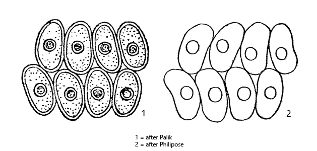
coenobia of 4–8–16 cells, arranged in 2 rows cells 7–18 µm long, width 3–9.9 µm
cell surface smooth
cells ovoid to cylindrical or curved (bean-shaped) with rounded apices
cells touch each other at their apices and longitudinal sides
with perforations between the apical contact points of the cells
one pyrenoid per cell
Cosmasiella arcuata var. platydisca
I have found Comasiella arcuata var. platydisca so far in the Bussenried as well as in the Simmelried. For identification it is important that the cells are staggered in two rows by half a cell width and that there is a small space between the contact points in the midline of the colony (s. fig. 1 a), where the cells are not in contact. In my population, the cells of the coenobia were almost always bean-shaped.
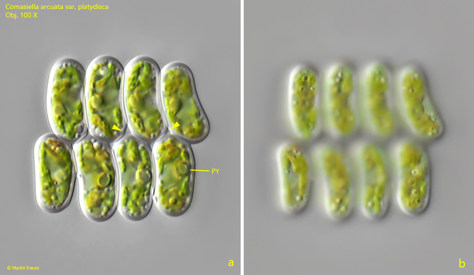
Fig. 1 a-b:Comasiella arcuata var. platydisca. L = 24 µm (of coenobium). Two focal planes of a coenobium of 8 cells. The cells are bean-shaped and 10–12 µm long (width 5 µm). The surface of the cells is smooth (b). Note the pore between the contact points of the apices of the cells (arrowheads). Obj. 100 X.