body broadly oval, almost quadrangular, dorso-ventrally flattened
length 45–70 µm
anterior end with a frontal plate
posterior end obliquely truncated
ventral side with a deep furrow running longitudinally
oral apparatus above cell equator
undulating membrane on left side of oral opening pouch-shaped
ciliation in three girdles (one anterior, two posterior)
ciliation interrupted in mid-body
macronucleus elliposoid, near mid-body or anterior third
one micronucleus adjacent to the macronucleus
one caudal cilium, inserted on right side of the postoral furrow
one contractile vacuole in the posterior third on the left side of the furrow
Cristigera pleuronemoides
I find Cristigera pleuronemoides in the mud layer of decomposing leaves and plant material. The species is especially common in autum after leaf fall.
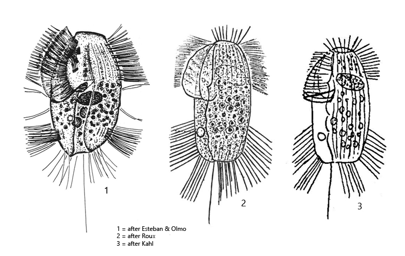
After the original description by Roux (1901) only few finds of Cristigera pleuronemoides are available. Kahl (1935) writes “bisher wohl noch nicht wiedergefunden (up to now probably not found again)” and obviously copies both drawing and description of Roux. Only in 1994 a re-description was published by Esteban & Olmo.
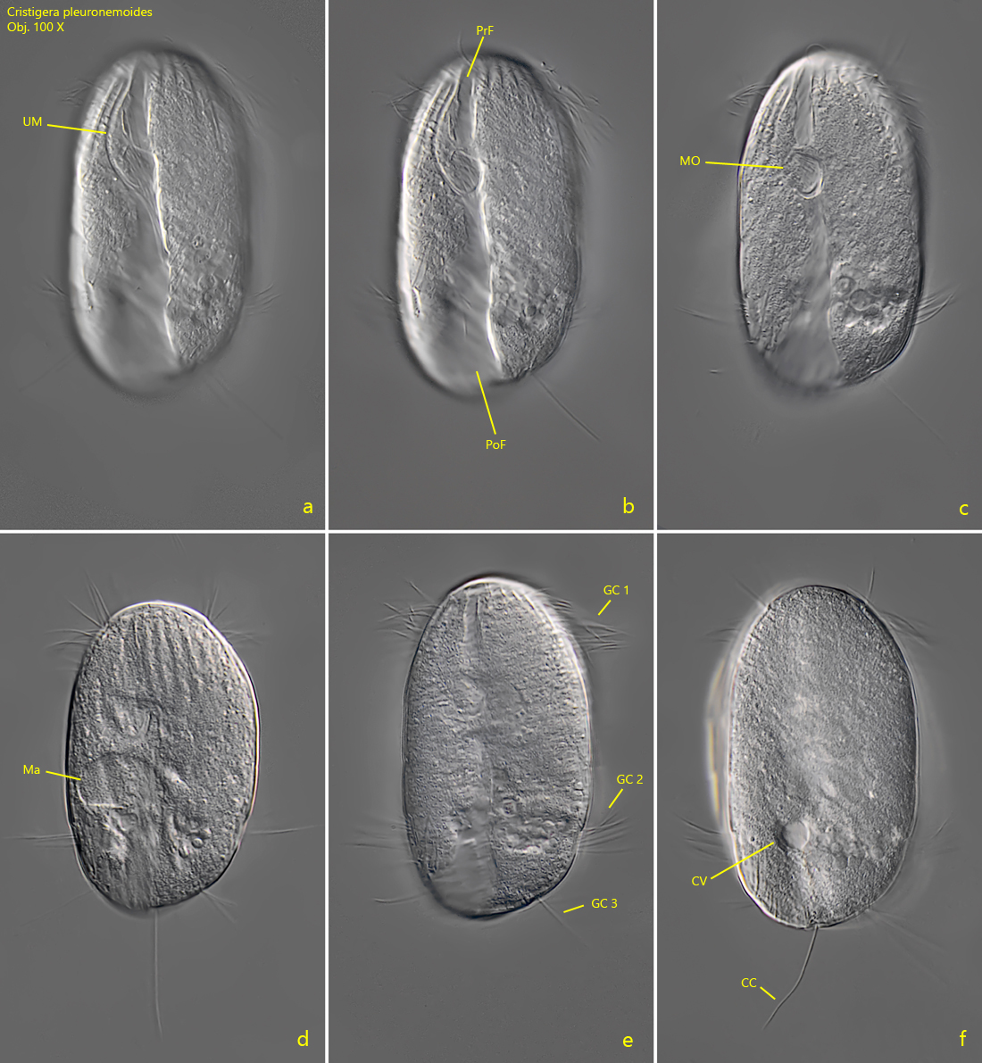
Cristigera pleuronemoides has a characteristic furrow that extends ventrally along the entire length of the body (s. fig. 2 a). The mouth opening is located in the anterior half of this furrow, with an undulating membrane on the left side. The mouth opening divides the furrow into a preoral and a postoral section (s. fig. 2 b). Another characteristic feature of Cristigera pleuronemoides are three separated girdles of cilia (s. figs. 2 e and 4 b). The first one is located at the anterior end and two others in the posterior third, while the mid-body is unciliated. In my population, all specimens examined had these three girdles of cilia. In the Spanish population described by Esteban & Olmer, there should be two additional girdles of cilia above and below the naked cell equator (s. drawing 1, above). I could not verify these additional girdles of cilia on any specimen of my population. While Roux and Kahl draw the posterior end straight truncated (s. drawings 2 and 3, above), in my population it was always obliquely truncated as drawn by Esteban & Olmer (s. drawing 1 , above). The macronucleus in my specimens was mostly broadly oval and located approximately in the middle of the body. All other characteristics agree with the descriptions of the above mentioned authors.
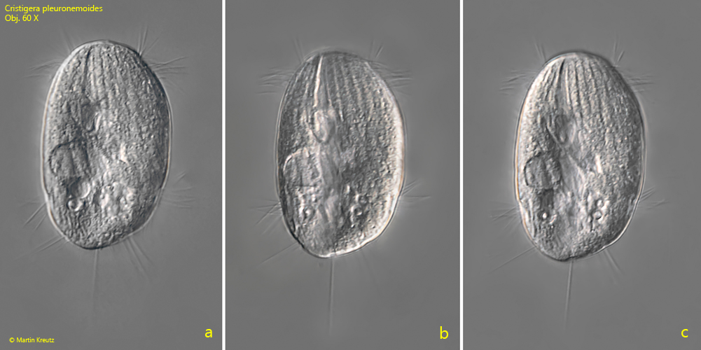
Fig. 1 a-c:Cristigera pleuronemoides. L = 65 µm. A freely swimming specimen from ventral. Obj. 60 X.
Fig. 2 a-f:Cristigera pleuronemoides. L = 68 µm. Different focal planes of a freely swimming specimen fom ventral. Note the three girdles of cilia (GC 1-3, e)and the longitudinal furrow separated by the mouth opening (MO) into a praeoral (PrF) and a postoral part (PoF). CC = caudal cilium, CV = contractile vacuole, Ma = macronucleus, UM = undulating membrane. Obj. 100 X
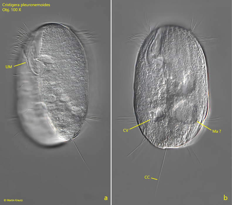
Fig. 3 a-b:Cristigera pleuronemoides. L = 70 µm. A third specimen from ventral. CC = caudal cilium, CV = contractile vacuole, Ma ? = probably the macronucleus. UM = undulating membrane. Obj. 100 X
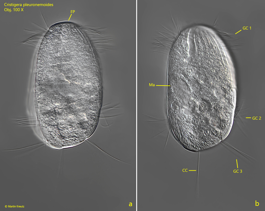
Fig. 4 a-b:Cristigera pleuronemoides. L = 65 µm. A fourth specimen from ventral. CC = caudal cilium, FP = frontal plate, GC 1-3 = girdles of cilia, Ma = macronucleus. Obj. 100 X